. 2017 Aug 15;8(1):247.
doi: 10.1038/s41467-017-00305-0.

Human microcephaly protein RTTN interacts with STIL and is required to build full-length centrioles

Affiliations

Human microcephaly protein RTTN interacts with STIL and is required to build full-length centrioles

Hsin-Yi Chen et al. Nat Commun. .

Abstract

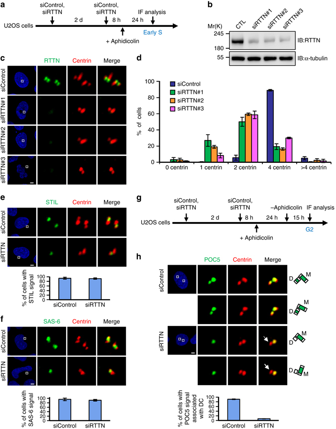
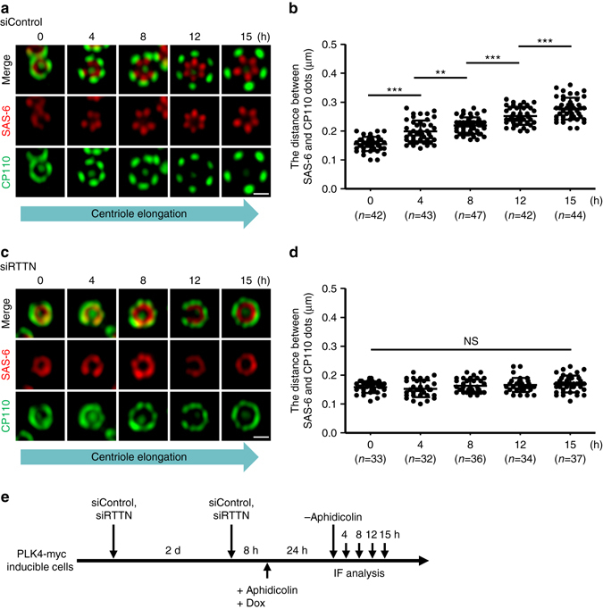
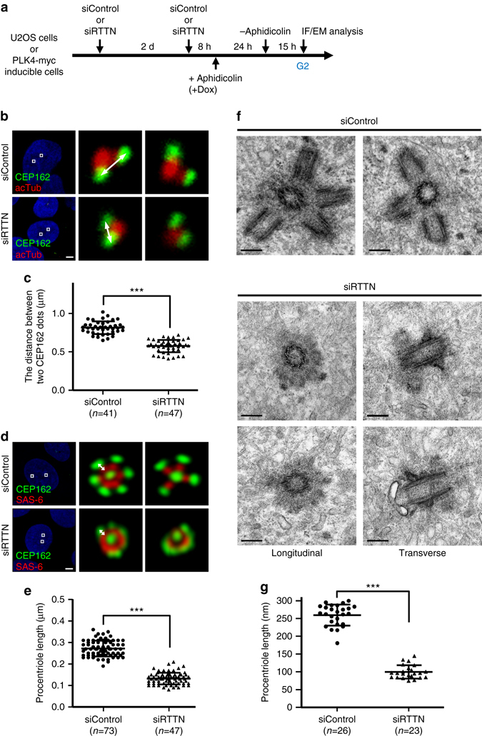
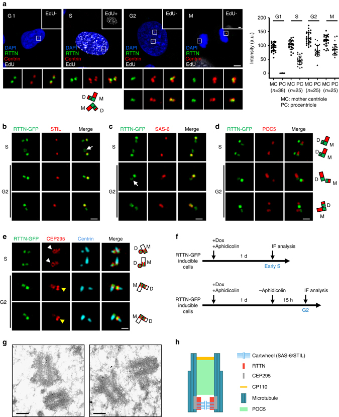
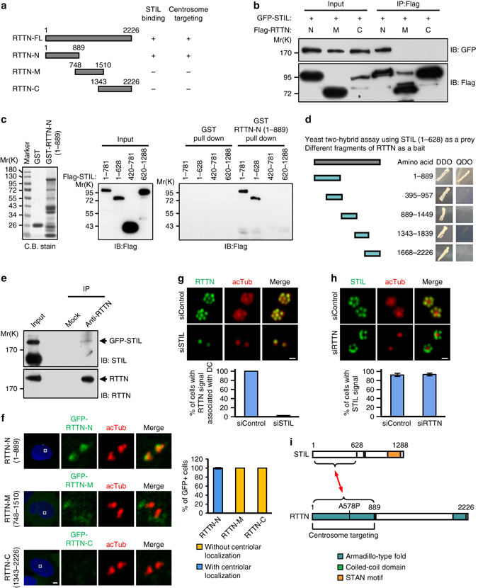
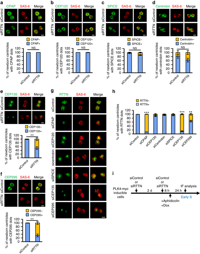
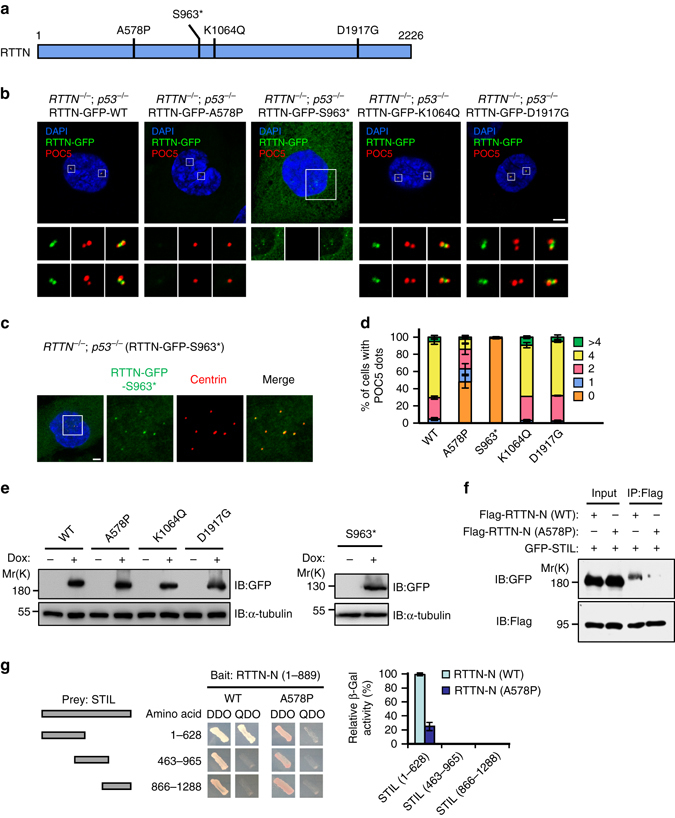
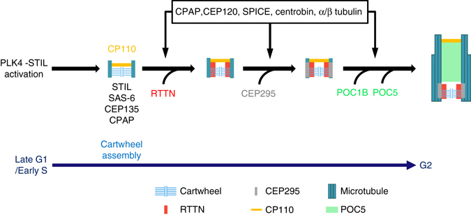
Mutations in many centriolar protein-encoding genes cause primary microcephaly. Using super-resolution and electron microscopy, we find that the human microcephaly protein, RTTN, is recruited to the proximal end of the procentriole at early S phase, and is located at the inner luminal walls of centrioles. Further studies demonstrate that RTTN directly interacts with STIL and acts downstream of STIL-mediated centriole assembly. CRISPR/Cas9-mediated RTTN gene knockout in p53-deficient cells induce amplification of primitive procentriole bodies that lack the distal-half centriolar proteins, POC5 and POC1B. Additional analyses show that RTTN serves as an upstream effector of CEP295, which mediates the loading of POC1B and POC5 to the distal-half centrioles. Interestingly, the naturally occurring microcephaly-associated mutant, RTTN (A578P), shows a low affinity for STIL binding and blocks centriole assembly. These findings reveal that RTTN contributes to building full-length centrioles and illuminate the molecular mechanism through which the RTTN (A578P) mutation causes primary microcephaly.Mutations in many centriolar protein-encoding genes cause primary microcephaly. Here the authors show that human microcephaly protein RTTN directly interacts with STIL and acts downstream of STIL-mediated centriole assembly, contributing to building full-length centrioles.


Conflict of interest statement

The authors declare no competing financial interests.

Figures

Fig. 1
Fig. 1
RTTN is required for normal centriole duplication. ac U2OS cells were treated with siControl or siRTTN (#1, #2, #3) and synchronized at early S phase, as described in a. The cells were analyzed by immunoblotting b or immunofluorescence confocal microscopy c using the indicated antibodies. d Histogram illustrating the percentages of cells exhibiting centrin signals. Error bars represent the mean ± s.d. from three independent experiments (n = 100/experiment). e, f U2OS cells were treated with siControl or siRTTN as described in a, and then stained with the indicated antibodies. Histogram illustrating the percentages of cells exhibiting STIL e or SAS-6 f signals. Error bars represent the mean ± s.d. from three independent experiments (n = 100/experiment). g, h U2OS cells were treated with siControl or siRTTN, enriched at G2 phase as described in g, and stained with the indicated antibodies. Histogram illustrating the percentages of cells with a POC5 signal associated with daughter centriole (DC). Error bars represent the mean ± s.d. from three independent experiments (n = 100/experiment). Scale bar, 5 μm
Fig. 2
Fig. 2
Depletion of RTTN perturbs centriole elongation. PLK4-myc-inducible cells were treated with siControl a, b or siRTTN c, d as shown in e, and analyzed by confocal fluorescence microscopy using the indicated antibodies. The procentriole length was measured as the distance between the fluorescent peak intensity of SAS-6 (red) and CP110 (green) in siControl- a, b and siRTTN-treated cells c, d. Error bars represent the mean ± s.d.; **P < 0.01; ***P < 0.001; NS, not significant (two-tailed t-test). Scale bar, 0.5 μm
Fig. 3
Fig. 3
Depletion of RTTN produces shorter centrioles. a, b U2OS cells were treated with siControl or siRTTN as shown in a, and analyzed by confocal fluorescence microscopy using the indicated antibodies. c Histogram illustrating the distance between the CEP162-positive dots associated with a given pair of orthogonally oriented centrioles. Error bars represent the mean ± s.d.; ***P < 0.001 (two-tailed t-test). d, f PLK4-myc doxycycline (Dox) inducible cells were treated with siControl or siRTTN as shown in a, and then analyzed by confocal fluorescence microscopy using the indicated antibodies d, or by electron microscopy f. e, g Histogram illustrating the length of procentrioles in PLK4-myc-inducible cells, as analyzed by confocal microscopy e or electron microscopy g. Error bars represent the mean ± s.d.; ***P < 0.001 (two-tailed t-test). The procentriole length in b/c was measured as the distance between two CEP162 dots as described by Azimzadeh et al. The procentriole length in d/e was measured as the distance between the fluorescent peak intensity of SAS-6 (red) and CEP162 (green) in siControl and siRTTN-treated cells. Scale bar, 5 μm in b and d; Scale bar, 200 nm in f
Fig. 4
Fig. 4
Super-resolution (3D-SIM) and immunogold electron microscopic analysis of the localization of RTTN during centriole biogenesis. a The subcellular localization of RTTN during the cell cycle. U2OS cells at different cell cycle stages were subjected to EdU labeling (white) and immunofluorescence staining using antibodies against RTTN (green) and centrin (red). EdU-labeled nuclei (EdU+) indicate cells at S phase. DNA was counterstained with DAPI (blue). The fluorescence intensity of RTTN at centrioles was quantified by ZEN software. bf RTTN-GFP doxycycline (Dox)-inducible cells were treated as described in f, and stained with antibodies against STIL b, SAS-6 c, POC5 d, CEP295 e, and centrin e. The fluorescence images were analyzed by 3D-SIM. M, mother centriole; D, daughter centriole. g Immunogold EM analysis of RTTN localization. h A schematic showing the localization of RTTN relative to those of other centriolar proteins at a centriole. Scale bar, 5 μm in a; Scale bar, 0.5 μm in be; Scale bar, 200 nm in g
Fig. 5
Fig. 5
RTTN directly interacts with and acts downstream of STIL. a Schematic representations of full-length RTTN-FL (1–2226), RTTN-N (1–889), RTTN-M (748–1510) and RTTN-C (1343–2226). b Mapping the STIL-interacting domain in RTTN. HEK293T cells were co-transfected with GFP-STIL and various Flag-tagged RTTN constructs, and then analyzed by immunoprecipitation (IP) and subsequent immunoblotting (IB) using the indicated antibodies. c GST-pulldown assay. GST and GST-RTTN-N (1–889) recombinant proteins were affinity purified and used to pulldown the indicated proteins from lysates of HEK293T cells that had been transfected with various Flag-STIL constructs. d Yeast two-hybrid assay shows a direct interaction between truncated STIL (1–628, prey) and various portions of RTTN (bait). The positive interaction between STIL and RTTN was demonstrated by the growth of mating colonies on QDO plates. e Endogenous RTTN and GFP-STIL form a complex in vivo. HEK293T cells were transfected with GFP-STIL. Twenty-four hours after transfection, the cell lysates were immunoprecipitated with anti-RTTN antibodies and immunoblotted with anti-STIL and anti-RTTN antibodies. f Mapping the centrosome-targeting region of RTTN. U2OS cells were transiently transfected with various GFP-tagged truncated RTTN constructs. At 24 h post-transfection, the cells were fixed and stained for acetylated tubulin (acTub). g RTTN acts downstream of STIL. PLK4-myc-inducible cells were treated with siControl or siSTIL and synchronized at early S phase by aphidicolin treatment. The cells were then subjected to immunofluorescence staining using the indicated antibodies, and the results were quantified. h PLK4-myc-inducible cells were treated with siControl or siRTTN as described in g, and the results were quantified. The error bars in f, g, and h represent the mean ± s.d. from three independent experiments (n = 100/experiment). i Schematic of the interaction between RTTN and STIL. Scale bar, 5 μm in f; Scale bar, 0.5 μm in g and h
Fig. 6
Fig. 6
Delineation of the RTTN-mediated centriole elongation pathway. af PLK4-myc-inducible cells were treated with siControl or siRTTN as shown in i and analyzed by confocal fluorescence microscopy using antibodies against SAS-6 af, CPAP a, CEP120 b, SPICE c, centrobin d, CEP135 e, or CEP295 f, and the results were quantified. Error bars represent the mean ± s.d. (n = 3 independent experiments with 100 cells scored per experiment). g PLK4-myc-inducible cells were treated with siRNAs against CPAP, CEP120, SPICE, centrobin, CEP135, or CEP295 as shown in i, and analyzed by confocal fluorescence microscopy using the indicated antibodies. h Histogram illustrating the percentages of newborn centrioles with RTTN dots. Error bars represent the mean ± s.d. (n = 3 independent experiments with 100 cells scored per experiment). **P < 0.01; ***P < 0.001; NS, not significant (two-tailed t-test). i Schematic of the procedure used to analyze the recruitment of centriolar proteins in siRNA-treated PLK4-myc-inducible cells. Scale bar, 0.5 μm
Fig. 7
Fig. 7
The human microcephaly RTTN mutant, A578P, exhibits reduced binding to STIL and its expression perturbs centriole duplication. a Schematic representation of RTTN showing the positions of RTTN mutations that cause primary microcephaly. b, c RTTN −/−; p53 −/− RPE1 cells that expressed doxycycline-inducible RTTN-GFP wild-type or mutant proteins (A578P, S963*, K1064Q, or D1917G) were synchronized by aphidicolin treatment for 24 h, and then released in fresh medium for another 15 h to allow progression to G2 phase. The cells were fixed and stained with the indicated antibodies. d Histogram illustrating the percentages of cells exhibiting POC5 dots. Error bars represent the mean ± s.d. from three independent experiments (n = 100/experiment). e RTTN −/−; p53 −/− RPE1 cells that expressed doxycycline-inducible RTTN-GFP wild-type or mutant proteins were analyzed by IB using the indicated antibodies. f The naturally occurring A578P mutation of RTTN reduces the binding of the protein to STIL. HEK293T cells were co-transfected Flag-RTTN-N (WT) or Flag-RTTN-N (A578P) mutant with full-length GFP-STIL. Twenty-four hours after transfection, cell lysates were IP with anti-Flag and analyzed by IB using the indicated antibodies. g Yeast two-hybrid assay testing interactions between various portions of STIL (prey) and truncated RTTN-N (bait, both WT and A578P). Histogram illustrating relative β-galactosidase (β-Gal) activity obtained in a liquid assay. The interaction between STIL (1–628) and RTTN-N (WT) was arbitrarily set to 100%. Error bars represent the mean ± s.d. from three independent experiments. Scale bar, 5 μm
Fig. 8
Fig. 8
Model showing the role of RTTN in procentriole elongation. During early S phase, RTTN is recruited to the inner luminal wall of newborn centrioles, where it likely stabilizes and maintains the primitive procentrioles. RTTN then helps recruit CEP295, which mediates procentriole elongation and promotes the loading of POC5/POC1B to the distal-half centrioles. CPAP and CEP120 promote the assembly of outer 9-triplet microtubules. For details, see the Discussion. For simplicity, only one centriole is shown

References

    1. Azimzadeh J, Marshall WF. Building the centriole. Curr. Biol. 2010;20:R816–R825. doi: 10.1016/j.cub.2010.08.010. - DOI - PMC - PubMed
    1. Gonczy P. Towards a molecular architecture of centriole assembly. Nat. Rev. Mol. Cell Biol. 2012;13:425–435. doi: 10.1038/nrm3373. - DOI - PubMed
    1. Conduit PT, Wainman A, Raff JW. Centrosome function and assembly in animal cells. Nat. Rev. Mol. Cell Biol. 2015;16:611–624. doi: 10.1038/nrm4062. - DOI - PubMed
    1. Habedanck R, Stierhof YD, Wilkinson CJ, Nigg EA. The polo kinase Plk4 functions in centriole duplication. Nat. Cell Biol. 2005;7:1140–1146. doi: 10.1038/ncb1320. - DOI - PubMed
    1. Kleylein-Sohn J, et al. Plk4-induced centriole biogenesis in human cells. Dev. Cell. 2007;13:190–202. doi: 10.1016/j.devcel.2007.07.002. - DOI - PubMed

MeSH terms