. 2017 Aug 15;7(1):8268.
doi: 10.1038/s41598-017-08583-w.

Inhibition of connexin hemichannels alleviates non-alcoholic steatohepatitis in mice

Affiliations

Inhibition of connexin hemichannels alleviates non-alcoholic steatohepatitis in mice

Joost Willebrords et al. Sci Rep. .

Abstract

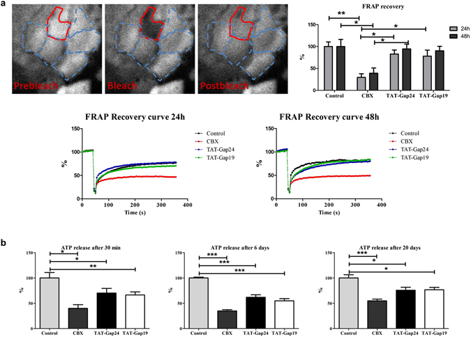
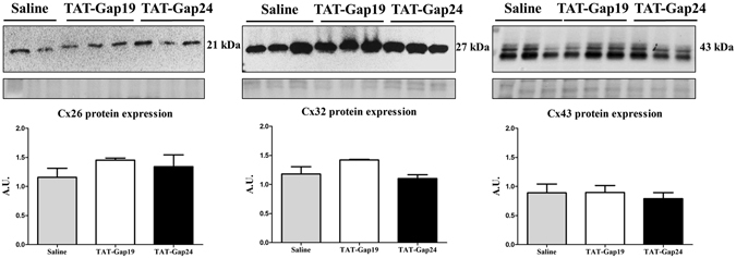
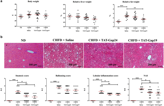
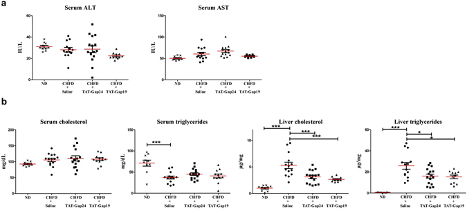
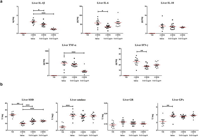
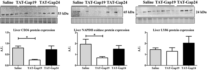
While gap junctions mediate intercellular communication and support liver homeostasis, connexin hemichannels are preferentially opened by pathological stimuli, including inflammation and oxidative stress. The latter are essential features of non-alcoholic steatohepatitis. In this study, it was investigated whether connexin32 and connexin43 hemichannels play a role in non-alcoholic steatohepatitis. Mice were fed a choline-deficient high-fat diet or normal diet for 8 weeks. Thereafter, TAT-Gap24 or TAT-Gap19, specific inhibitors of hemichannels composed of connexin32 and connexin43, respectively, were administered for 2 weeks. Subsequently, histopathological examination was carried out and various indicators of inflammation, liver damage and oxidative stress were tested. In addition, whole transcriptome microarray analysis of liver tissue was performed. Channel specificity of TAT-Gap24 and TAT-Gap19 was examined in vitro by fluorescence recovery after photobleaching analysis and measurement of extracellular release of adenosine triphosphate. TAT-Gap24 and TAT-Gap19 were shown to be hemichannel-specific in cultured primary hepatocytes. Diet-fed animals treated with TAT-Gap24 or TAT-Gap19 displayed decreased amounts of liver lipids and inflammatory markers, and augmented levels of superoxide dismutase, which was supported by the microarray results. These findings show the involvement of connexin32 and connexin43 hemichannels in non-alcoholic steatohepatitis and, simultaneously, suggest a role as potential drug targets in non-alcoholic steatohepatitis.


Conflict of interest statement

The authors declare that they have no competing interests.

Figures

Figure 1
Figure 1
Effects of TAT-Gap24 and TAT-Gap19 on gap junction activity and connexin hemichannel responses. Primary rat hepatocytes were exposed to 50 µM CBX, 20 µM TAT-Gap24, 20 µM TAT-Gap19 or vehicle control. (a) Gap junction activity measured through FRAP analysis after 24 h and 48 h (n = 4, N = 4). Fluorescence in the bleached cell was expressed as the percentage of recovery relative to the starting level just before photobleaching. A similar analysis was performed on cells distant from the bleached area to monitor photobleaching outside the targeted region. The recovery values for each experiment were normalized to the corresponding vehicle control condition. Pre-bleach (left panel), bleach (middle panel), post-bleach (right panel) after 6 min. (b) ATP release 30 min after incubation of TAT-Gap24, TAT-Gap19 or vehicle control for 0 min, 6 days and 20 days (n = 3, N = 6). Data are expressed as means ± SEM with *p < 0.5, **p < 0.01 and ***p < 0.001.
Figure 2
Figure 2
Effects of TAT-Gap24 and TAT-Gap19 on connexin protein expression in liver. After 8 weeks of CHFD, an osmotic pump was surgically implanted in the abdominal cavity, which ensured sustained release of 1 mg/kg/day TAT-Gap24 (n = 11) or TAT-Gap19 (n = 12) or saline (n = 14) for another 2 weeks, while continuing the diet. Immunoblot analysis of Cx26 (21 kDa), Cx32 (27 kDa) and Cx43 (43 kDa) after separation and blotting, after which results were normalized to total protein loading. Blot images are cropped. Full-length blots are presented in Supplementary Fig. 4. Data are expressed as means ± SEM.
Figure 3
Figure 3
Effects of TAT-Gap24 and TAT-Gap19 on biometric parameters and liver histology in NASH. After 8 weeks of CHFD, an osmotic pump was surgically implanted in the abdominal cavity, which ensured sustained release of 1 mg/kg/day TAT-Gap24 (n = 11) or TAT-Gap19 (n = 12) or saline (n = 14) for another 2 weeks while continuing the diet. (a) Body, fat and liver of mice and relative liver and fat weight. (b) Steatosis, lobular inflammation, ballooning and NAS score based on hematoxylin-eosin staining of liver tissue of ND group (first panel), saline group (second panel), TAT-Gap24 (third panel) and TAT-Gap19 group (fourth panel). Data are expressed as means ± SEM with *p < 0.5, **p < 0.01 and ***p < 0.001.
Figure 4
Figure 4
Effects of TAT-Gap24 and TAT-Gap19 on serum transaminases and serum and liver lipid content in NASH. After 8 weeks of CHFD, an osmotic pump was surgically implanted in the abdominal cavity, which ensured sustained release of 1 mg/kg/day TAT-Gap24 (n = 11) or TAT-Gap19 (n = 11) or saline (n = 13) for another 2 weeks while continuing the diet. (a) Serum ALT and AST. (b) Serum and liver triglycerides and cholesterol. Data are expressed as means ± SEM with *p < 0.05 and ***p < 0.001.
Figure 5
Figure 5
Effects of TAT-Gap24 and TAT-Gap19 on inflammatory cytokines and oxidative stress in NASH. After 8 weeks of CHFD, an osmotic pump was surgically implanted in the abdominal cavity, which ensured sustained release of 1 mg/kg/day TAT-Gap24 (n = 11) or TAT-Gap19 (n = 12) or saline (n = 14) for another 2 weeks while continuing the diet. (a) Levels of IFN-γ, IL-6, IL-1β, TNF-α and IL-10 in liver tissue and serum. (b) Activity of SOD, GR, GPx and catalase in liver tissue. Data are expressed as means ± SEM with *p < 0.05, **p < 0.01, and ***p < 0.001.
Figure 6
Figure 6
Protein expression analysis of liver tissue of TAT-Gap24 and TAT-Gap19-treated NASH animals. After 8 weeks of CHFD, an osmotic pump was surgically implanted in the abdominal cavity, which ensured sustained release of 1 mg/kg/day TAT-Gap24 (n = 6) or TAT-Gap19 (n = 8) or saline (n = 9) for another 2 weeks while continuing the diet. Immunoblot analysis of CD36 (53 kDa), NADPH oxidase (65 kDa) and LY86 (24 kDa) after separation and blotting, after which results were normalized to total protein. Blot images are cropped. Full-length blots are presented in Supplementary Fig. 5. Data are expressed as means ± SEM with *p < 0.05.

References

    1. Byrne CD, Targher G. NAFLD: a multisystem disease. J Hepatol. 2015;62:S47–64. doi: 10.1016/j.jhep.2014.12.012. - DOI - PubMed
    1. Carr RM, Oranu A, Khungar V. Nonalcoholic Fatty Liver Disease: Pathophysiology and Management. Gastroenterol Clin North Am. 2016;45:639–652. doi: 10.1016/j.gtc.2016.07.003. - DOI - PMC - PubMed
    1. Willebrords J, et al. Strategies, models and biomarkers in experimental non-alcoholic fatty liver disease research. Prog Lipid Res. 2015;59:106–125. doi: 10.1016/j.plipres.2015.05.002. - DOI - PMC - PubMed
    1. Buzzetti E, Pinzani M, Tsochatzis EA. The multiple-hit pathogenesis of non-alcoholic fatty liver disease (NAFLD) Metabolism. 2016;65:1038–1048. doi: 10.1016/j.metabol.2015.12.012. - DOI - PubMed
    1. Peverill W, Powell LW, Skoien R. Evolving concepts in the pathogenesis of NASH: beyond steatosis and inflammation. Int J Mol Sci. 2014;15:8591–8638. doi: 10.3390/ijms15058591. - DOI - PMC - PubMed

Publication types

MeSH terms