. 2017 Aug 15;7(1):8197.
doi: 10.1038/s41598-017-08572-z.

Mussel Inspired Polynorepinephrine Functionalized Electrospun Polycaprolactone Microfibers for Muscle Regeneration

Affiliations

Mussel Inspired Polynorepinephrine Functionalized Electrospun Polycaprolactone Microfibers for Muscle Regeneration

Ying Liu et al. Sci Rep. .

Abstract

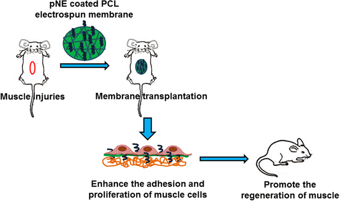
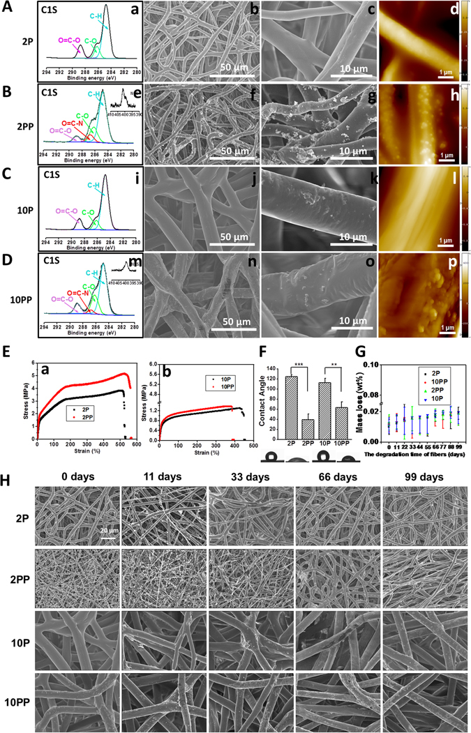
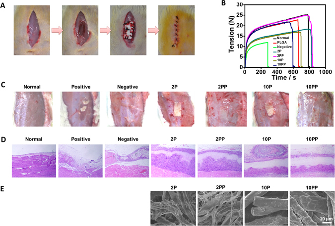
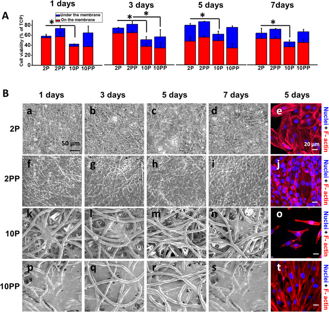
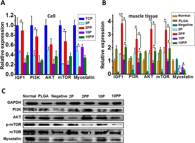
Electrospun scaffolds with excellent mechanical properties, high specific surface area and a commendable porous network are widely used in tissue engineering. Improving the hydrophilicity and cell adhesion of hydrophobic substrates is the key point to enhance the effectiveness of electrospun scaffolds. In this study, polycaprolactone (PCL) fibrous membranes with appropriate diameter were selected and coated by mussel-inspired poly norepinephrine (pNE). And norepinephrine is a catecholamine functioning as a hormone and neurotransmitter in the human brain. The membrane with smaller diameter fibers, a relative larger specific surface area and the suitable pNE functionalization provided more suitable microenvironment for cell adhesion and proliferation both in vitro and in vivo. The regenerated muscle layer can be integrated well with fibrous membranes and surrounding tissues at the impaired site and thus the mechanical strength reached the value of native tissue. The underlying molecular mechanism is mediated via inhibiting myostatin expression by PI3K/AKT/mTOR hypertrophy pathway. The properly functionalized fibrous membranes hold the potential for repairing muscle injuries. Our current work also provides an insight for rational design and development of better tissue engineering materials for skeletal muscle regeneration.


Conflict of interest statement

The authors declare that they have no competing interests.

Figures

Figure 1
Figure 1
Scheme of pNE coated PCL electrospun fibrous membranes and their effect on muscle regeneration in vitro and in vivo.
Figure 2
Figure 2
Characterization of PCL fibrous membranes. (AD) XPS spectra (a,e,i,m), SEM (b,c,f,g,j,k,n,o) and AFM images (d,h,l,p). A, PCL fibrous membrane with 2 μm in diameter (2 P). B, pNE coated PCL fibrous membrane with 2 μm in diameter (2 PP). C, PCL fibrous membrane with 10 μm in diameter (10 P). D, pNE coated PCL fibrous membrane with 10 μm in diameter (10 PP). (E-H) The mechanical property, wettability and degradation in vitro of PCL fibrous membranes. E, The stress-strain curves of PCL fibrous membranes. F, Images and quantitative statistics of contact angle. G, Mass loss of PCL fibrous membranes after cultured in PBS at 37 °C for 11, 33, 66 and 99 days, respectively. H, SEM images of PCL fibrous membranes after cultured in PBS at 37 °C for 11, 33, 66 and 99 days.
Figure 3
Figure 3
Functionalized electrospun PCL memberanes were effective for the regenerating muscle injury. (A) Diagram of PCL fibrous membranes implantation in vivo. After implantation into rat muscles for 40 days, mechanical properties of PCL fibrous membranes (B), visible morphology (C) and histological observations (D) of muscles and SEM images of PCL fibrous membranes (E) were observed.
Figure 4
Figure 4
Thinner functionalized electrospun PCL microfibers (2 PP) were more effective for the muscle cell proliferation and adhesion. (A) After cultured on PCL fibrous membranes for 1, 3, 5 and 7 days, cell proliferation was detected using CCK-8. (B) ESEM and fluorescent images of cells on PCL fibrous membranes.
Figure 5
Figure 5
Thinner functionalized electrospun PCL microfibers (2 PP) were more effective for the muscle cell growth and differentiation. (A,B) Relative expression of IGF1, PI3K, AKT, mTOR and myostatin of cells and muscle tissues. C, Representative western blotting of PI3K, AKT and myostatin of muscle tissues from in vivo experiments. *, #, $ p < 0.05 and **p < 0.01 as indicated.

References

    1. Bao M, et al. Electrospun biomimetic fibrous scaffold from shape memory polymer of PDLLA-co-TMC for bone tissue engineering. ACS Appl. Mater. Inter. 2014;6(4):2611–2621. doi: 10.1021/am405101k. - DOI - PubMed
    1. Brun P, et al. Electrospun scaffolds of self-assembling peptides with poly(ethylene oxide) for bone tissue engineering. Acta Biomater. 2011;7(6):2526–2532. doi: 10.1016/j.actbio.2011.02.025. - DOI - PubMed
    1. Shi X, Zhao Y, Zhou J, Chen S, Wu H. One-Step Generation of Engineered Drug-Laden Poly(lactic-co-glycolic acid) Micropatterned with Teflon Chips for Potential Application in Tendon Restoration. ACS Appl. Mater. Inter. 2013;5(21):10583–10590. doi: 10.1021/am402388k. - DOI - PubMed
    1. Chen B, et al. In vivo tendon engineering with skeletal muscle derived cells in a mouse model. Biomaterials. 2012;33(26):6086–6097. doi: 10.1016/j.biomaterials.2012.05.022. - DOI - PubMed
    1. Cittadella Vigodarzere G, Mantero S. Skeletal muscle tissue engineering: strategies for volumetric constructs. Front. Physiol. 2014;5 doi: 10.3389/fphys.2014.00362. - DOI - PMC - PubMed

Publication types