Review
. 2017 Oct;18(10):622-636.
doi: 10.1038/nrm.2017.67. Epub 2017 Aug 16.

The essential kinase ATR: ensuring faithful duplication of a challenging genome

Affiliations
Review

The essential kinase ATR: ensuring faithful duplication of a challenging genome

Joshua C Saldivar et al. Nat Rev Mol Cell Biol. 2017 Oct.

Erratum in

Abstract

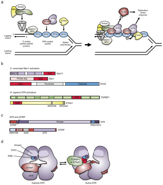
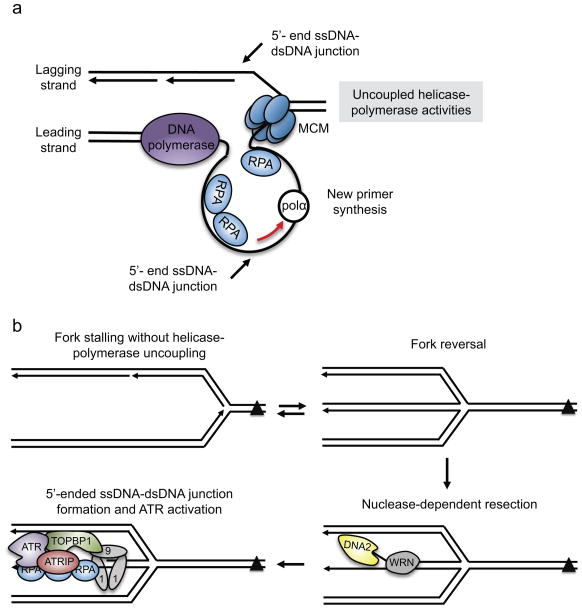
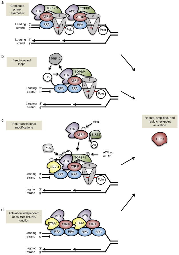
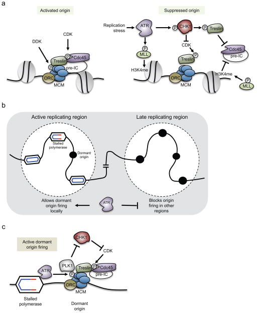
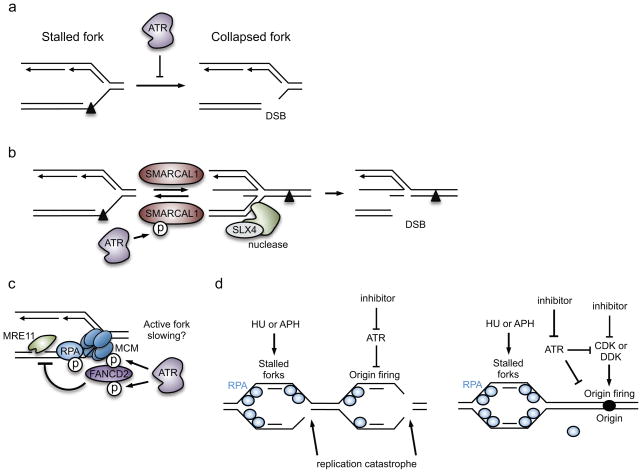
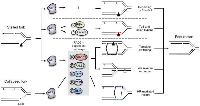
One way to preserve a rare book is to lock it away from all potential sources of damage. Of course, an inaccessible book is also of little use, and the paper and ink will continue to degrade with age in any case. Like a book, the information stored in our DNA needs to be read, but it is also subject to continuous assault and therefore needs to be protected. In this Review, we examine how the replication stress response that is controlled by the kinase ataxia telangiectasia and Rad3-related (ATR) senses and resolves threats to DNA integrity so that the DNA remains available to read in all of our cells. We discuss the multiple data that have revealed an elegant yet increasingly complex mechanism of ATR activation. This involves a core set of components that recruit ATR to stressed replication forks, stimulate kinase activity and amplify ATR signalling. We focus on the activities of ATR in the control of cell cycle checkpoints, origin firing and replication fork stability, and on how proper regulation of these processes is crucial to ensure faithful duplication of a challenging genome.


Conflict of interest statement

Competing interests statement

The authors declare no competing interests.

Figures

Figure 1
Figure 1. Components of ATR activation pathways
(a, Left) DNA polymerase stalling on the lagging strand generates a single-stranded DNA (ssDNA) gap that is bound by replication protein A (RPA), providing a platform for ataxia telangiectasia and Rad3-related (ATR) activation. The 5′-ended ssDNA–dsDNA junction formed at the Okazaki fragment adjacent to this ssDNA serves as the loading point for the RAD9–RAD1–HUS1 (9-1-1) clamp complex, which is loaded onto the DNA by RAD17–replication factor C subunits 2-5 (RFC2-5) clamp loader. (a, Right) The 9-1-1 complex with assistance from RHINO (RAD9–HUS1–RAD1 interacting nuclear orphan 1) and the MRE11–RAD50–NBS1 (MRN) complex recruits the ATR activator topoisomerase II binding protein (TOPBP1), thereby allowing stimulation of ATR and phosphorylation of specific downstream effectors, including checkpoint kinase 1 (CHK1). Ewings tumor associated antigen (ETAA1) bound to RPA activates ATR in a parallel pathway. (b) In budding yeast Dpb11, DNA damage checkpoint protein 1 (Ddc1) and Dna2 have a disordered ATR activating domain (AAD), which contains critical hydrophobic amino acids that are needed for Mec1 (ATR homologue) activation. In humans, the ATR activator TOPBP1 is a homolog of Dpb11, whereas ETAA1 is not related to any yeast protein, and unlike the other activators, contains two motifs (RPA70N and RPA32C) that interact with two domains of RPA. (c) The ATR kinase domain is followed by two motifs that are needed for ATR activation: FATC, and PIKK-regulatory domain (PRD), which may directly contact the AAD. ATR-interacting protein (ATRIP) contains an amino-terminal RPA-interaction domain (RPA70N), a coiled-coil dimerization domain (CC), a motif that is needed for AAD interaction and ATR activation, and a carboxy-terminal region that interacts with ATR,–. (d) Model of ATR activation. ATR–ATRIP forms a dimer of dimers. TOPBP1 or ETAA1 AAD binding likely induces a conformational change in ATR that reduces the Km of ATR for its substrates and thereby activates ATR. BRCT, BRCA1 C Terminus.
Figure 2
Figure 2. Generation of the ATR-activating structure at stressed replication forks
(a) When the leading strand polymerase stalls, a 5′-ended ssDNA–dsDNA junction is not initially present. New primer synthesis ahead of the stalled leading-strand polymerase would create the ssDNA–dsDNA junction. DNA polymerase alpha (Pol α) and/or primase and DNA directed polymerase (PrimPol) (not shown) may catalyze primer synthesis ahead of the stalled polymerase. (b) Fork remodeling may be necessary to generate the ATR-activating structure when a DNA lesion, such as an inter-strand crosslink (ICL), stalls the fork entirely. In this situation, DNA translocases may reverse the fork. Once reversed, specialized helicases, such as Werner syndrome RecQ like helicase (WRN) or Fanconi anemia complementation group J (FANCJ) (not shown), can unwind the dsDNA of the reversed strands. Exonucleases, including DNA replication helicase/nuclease 2 (DNA2), can resect the DNA in the 5′-3′ direction as it is unwound by the helicase, generating both the ssDNA and the 5′-ended ssDNA–dsDNA junction required for ATR activation. MCM2-7, minichromosome maintenance 2-7 complex; RPA, Replication protein A; ATRIP, ATR-interacting protein; TOBP1, topoisomerase II binding protein 1; 9-1-1, RAD9–RAD1–HUS1
Figure 3
Figure 3. Amplification of ATR signaling
There are multiple ways to amplify ATR signaling at individual replication forks. (a) Continued primer synthesis by DNA polymerase alpha (Pol α) ahead of the stalled leading-strand polymerase can generate multiple ssDNA–dsDNA junctions at a single fork. This would create multiple loading points for the RAD9–RAD1–HUS1 (9-1-1) complex and the ATR activator topoisomerase II binding protein 1 (TOPBP1), and, accordingly, would increase the number of ATR proteins that are activated at a single fork. (b) The E3 ubiquitin ligase PRP19 creates a feed-forward loop to amplify ATR activity. PRP19 ubiquitylates replication protein A (RPA), which increases ATR activity, which in turn boosts PRP19-dependent ubiquitylation of RPA. (c) Multiple post-translational modifications amplify ATR activation. These include protein kinase A (PKA) phosphorylation of ATR, ATR autophosphorylation, cyclin-dependent kinase 2 (CDK2) phosphorylation of ATR-interacting protein (ATRIP), ATR and/or ATM phosphorylation of TOPBP1, and potentially ATR phosphorylation of Ewing tumor-associated antigen 1 (ETAA1). ATRIP deacetylation by Sirtuin 2 (SIRT2) helps recruit ATR–ATRIP to the stalled replication fork. (d) The second ATR activator, ETAA1, can stimulate ATR activity independently of the presence of ssDNA–dsDNA junctions. Because ETAA1, ATR and ATRIP are recruited to RPA–ssDNA, longer stretches of ssDNA could recruit multiple ETAA1, ATR and ATRIP proteins to a single fork and produce an amplified ATR-mediated response. CHK1, checkpoint kinase 1
Figure 4
Figure 4. Pathways regulated by ATR to suppress origin firing
(a) (Left) DBF4-dependent kinase (DDK) and cyclin-dependent kinase (CDK) activities promote origin firing. DDK phosphorylates the minichromosome maintenance 2-7 complex helicase (MCM), and CDK phosphorylates Treslin. These phosphorylations promote recruitment of cell division cycle 45 (CDC45) and other pre-initiation complex (pre-IC) factors to activate the helicase.(Right) ATR may suppress origin firing through at least two distinct pathways. The first is through phosphorylation and stabilization of myeloid/lymphoid or mixed-lineage leukemia (MLL). This promotes MLL association with chromatin where it methylates histone H3 Lys4 (H3K4me). This chromatin modification blocks CDC45 loading onto nearby origins. The second pathway is through ATR-dependent activation of checkpoint kinase 1 (CHK1), which negatively regulates CDK-dependent phosphorylations at origins, thereby blocking the loading of CDC45 and other pre-IC factors. CHK1 also directly phosphorylates Treslin, which limits CDC45 binding to origins. (b and c) ATR allows local dormant origins to fire in response to replication stress. (b) When a replication fork is stalled, nearby dormant origins fire to help complete DNA synthesis in its vicinity. At the same time, cells also block origin firing in later replicating regions. This prevents the accumulation of additional replication stress and the potential depletion of replication factors or nucleotides. (c) It is unclear how ATR allows dormant origins to fire locally. One proposed mechanism involves inhibition of CHK1 activity in the vicinity of the stalled polymerase. ATR activated at the stalled fork can phosphorylates MCM2 locally, primarily at nearby unfired origins. MCM2 phosphorylation creates a docking site for polo-like kinase 1 (PLK1), which suppresses activation of CHK1 and allows recruitment of CDC45 to nearby origins. ORC, origin recognition complex
Figure 5
Figure 5. Proposed mechanisms by which ATR maintains replication-fork stability
(a) ATR prevents fork collapse, which is illustrated here as double-strand DNA break (DSB) formation at the fork. In this example, replication of the leading strand was blocked by a DNA lesion. (b) ATR phosphorylates SWI/SNF related, matrix associated, actin dependent regulator of chromatin, subfamily a like 1 (SMARCAL1) at stressed replication forks, thereby suppressing its activity and limiting fork reversal. As structure-specific endonuclease subunit (SLX4)-dependent nucleases can cleave reversed forks, ATR-dependent inhibition of SMARCAL1 is a mechanism by which ATR stabilizes the fork. (c) ATR phosphorylates several proteins at the replication fork to modify replisome function. ATR mediates the recruitment of Fanconi anemia complementation group D2 (FANCD2) to the fork, which may occur in response to ATR-dependent phosphorylation of the MCM2-7 helicase and FANCD2. FANCD2 minimizes the accumulation of ssDNA caused by meiotic recombination 11 homolog A (MRE11)-dependent resection of the nascent DNA. FANCD2 also slows DNA polymerases at stressed forks, and fork slowing may prevent collapse. (d) ATR prevents exhaustion of replication protein A (RPA) availability and subsequently replication catastrophe. This indirect function of ATR is mediated partly through its role in suppressing origin firing in response to hydroxyurea (HU)- or aphidicolin (APH)-induced replication stress.
Figure 6
Figure 6. Proposed roles of ATR in promoting replication-fork restart
In addition to stabilizing the replication fork, ATR is thought to promote restart of stalled forks. There are several pathways to restart a stalled fork, including repriming ahead of the lesion by primase and DNA directed polymerase (PrimPol), lesion bypass through translesion synthesis (TLS), lesion bypass through template switching, and fork reversal and lesion repair. If a stalled fork collapses into a double-strand DNA break (DSB), homologous recombination-dependent pathways can restart the fork. It is unknown whether ATR regulates PrimPol activity to restart replication forks, but ATR does phosphorylate two TLS polymerases, REV1 and DNA polymerase eta (Pol η), and may promote lesion bypass. ATR also phosphorylates several proteins that promote radiation sensitive 51 (RAD51)-dependent replication restart pathways, including template switching, fork reversal and repair, and homologous recombination. These include X-ray repair cross complementing 3 (XRCC3), partner and localizer of BRCA2 (PALB2), replication protein A (RPA), Werner syndrome RecQ like helicase (WRN) and Bloom syndrome RecQ like helicase (BLM).

References

    1. Zeman MK, Cimprich KA. Causes and consequences of replication stress. Nature Cell Biol. 2014;16:2–9. - PMC - PubMed
    1. Brown EJ, Baltimore D. ATR disruption leads to chromosomal fragmentation and early embryonic lethality. Genes Dev. 2000;14:397–402. Demonstrated that ATR is essential for viability in vivo. - PMC - PubMed
    1. de Klein A, et al. Targeted disruption of the cell-cycle checkpoint gene ATR leads to early embryonic lethality in mice. Curr Biol. 2000;10:479–482. Demonstrated that ATR is essential for viability in vivo. - PubMed
    1. O’Driscoll M, Ruiz-Perez VL, Woods CG, Jeggo PA, Goodship JA. A splicing mutation affecting expression of ataxia-telangiectasia and Rad3-related protein (ATR) results in Seckel syndrome. Nature Genet. 2003;33:497–501. - PubMed
    1. Cimprich KA, Shin TB, Keith CT, Schreiber SL. cDNA cloning and gene mapping of a candidate human cell cycle checkpoint protein. Proc Natl Acad Sci USA. 1996;93:2850–2855. - PMC - PubMed

Substances