

Development and characterization of naive single-type tumor antigen-specific CD8+ T lymphocytes from murine pluripotent stem cells
- PMID: 28811978
- PMCID: PMC5543906
- DOI: 10.1080/2162402X.2017.1334027
Development and characterization of naive single-type tumor antigen-specific CD8+ T lymphocytes from murine pluripotent stem cells
Abstract
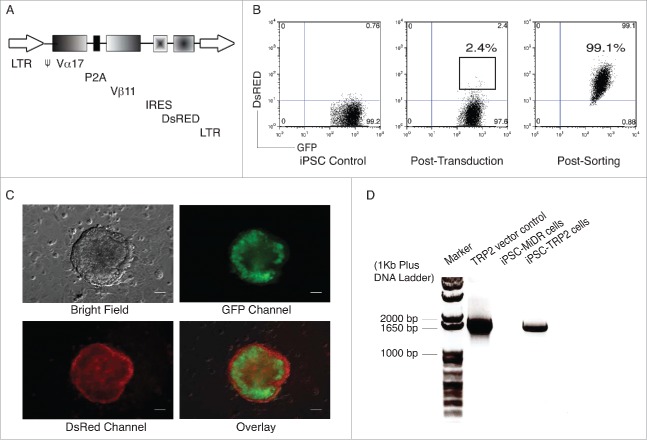
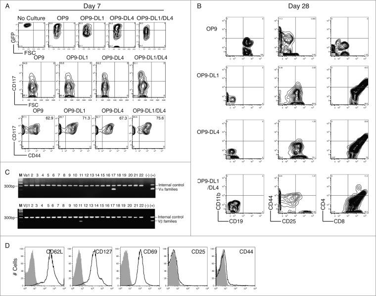
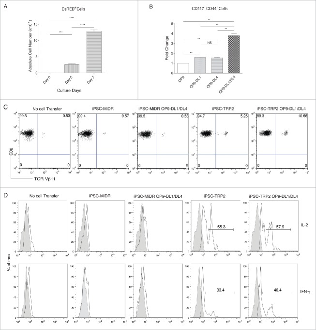
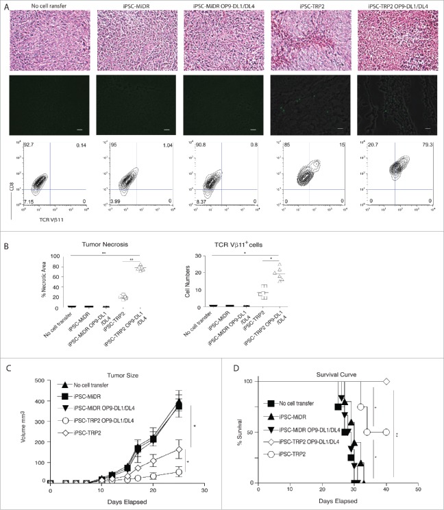
Optimal approaches to differentiate tumor antigen-specific cytotoxic T lymphocytes (CTLs) from pluripotent stem cells (PSCs) remain elusive. In the current study, we showed that combination of in vitro priming through Notch ligands and in vivo development facilitated the generation of tumor Ag-specific CTLs that effectively inhibited tumor growth. We co-cultured the murine induced PSCs (iPSCs) genetically modified with tyrosinase-related protein 2 (TRP2)-specific T cell receptors with OP9 cell line expressing both Notch ligands Delta-like 1 and 4 (OP9-DL1/DL4) for a week before adoptively transferred into recipient C67BL/6 mice. Three weeks later, B16 melanoma cells were inoculated subcutaneously, and the antitumor activity of the iPSC-derived T cells was assessed. We observed the development of the TRP2-specific iPSC-CD8+ T cells that responded to Ag stimulation and infiltrated into melanoma tissues, significantly inhibited the tumor growth, and improved the survival of the tumor-bearing mice. Thus, this approach may provide a novel effective strategy to treatment of malignant tumors.
Keywords: Cytotoxic T lymphocytes; Pluripotent stem cells; immunotherapy; melanoma, mouse.
Figures
Similar articles
-
Melanoma Immunotherapy in Mice Using Genetically Engineered Pluripotent Stem Cells.Cell Transplant. 2016;25(5):811-27. doi: 10.3727/096368916X690467. Epub 2016 Jan 15. Cell Transplant. 2016. PMID: 26777320 Free PMC article.
-
Protective Cancer Vaccine Using Genetically Modified Hematopoietic Stem Cells.Vaccines (Basel). 2018 Jul 6;6(3):40. doi: 10.3390/vaccines6030040. Vaccines (Basel). 2018. PMID: 29986440 Free PMC article.
-
In vivo programming of tumor antigen-specific T lymphocytes from pluripotent stem cells to promote cancer immunosurveillance.Cancer Res. 2011 Jul 15;71(14):4742-7. doi: 10.1158/0008-5472.CAN-11-0359. Epub 2011 May 31. Cancer Res. 2011. PMID: 21628492
-
Cloning and expansion of antigen-specific T cells using iPS cell technology: development of "off-the-shelf" T cells for the use in allogeneic transfusion settings.Int J Hematol. 2018 Mar;107(3):271-277. doi: 10.1007/s12185-018-2399-1. Epub 2018 Jan 31. Int J Hematol. 2018. PMID: 29388165 Review.
-
CD8+ cytotoxic T lymphocytes in cancer immunotherapy: A review.J Cell Physiol. 2019 Jun;234(6):8509-8521. doi: 10.1002/jcp.27782. Epub 2018 Nov 22. J Cell Physiol. 2019. PMID: 30520029 Review.
Cited by
-
Generation of Tumor Antigen-Specific Cytotoxic T Lymphocytes from Pluripotent Stem Cells.Methods Mol Biol. 2019;1884:43-55. doi: 10.1007/978-1-4939-8885-3_3. Methods Mol Biol. 2019. PMID: 30465194 Free PMC article.
-
Stem Cell-Derived Viral Antigen-Specific T Cells Suppress HBV Replication through Production of IFN-γ and TNF-⍺.iScience. 2020 Jul 24;23(7):101333. doi: 10.1016/j.isci.2020.101333. Epub 2020 Jul 1. iScience. 2020. PMID: 32679546 Free PMC article.
-
In Vitro Differentiation of T Cells from Murine Pluripotent Stem Cells.Methods Mol Biol. 2019;2048:131-141. doi: 10.1007/978-1-4939-9728-2_14. Methods Mol Biol. 2019. PMID: 31396937 Free PMC article.
-
Stem Cell-Derived Viral Ag-Specific T Lymphocytes Suppress HBV Replication in Mice.J Vis Exp. 2019 Sep 25;(151):10.3791/60043. doi: 10.3791/60043. J Vis Exp. 2019. PMID: 31609353 Free PMC article.
References
-
- Rosenberg SA, Restifo NP. Adoptive cell transfer as personalized immunotherapy for human cancer. Science 2015; 348:62-8; PMID:25838374; https://doi.org/10.1126/science.aaa4967 - DOI - PMC - PubMed
-
- Antsiferova O, Muller A, Ramer PC, Chijioke O, Chatterjee B, Raykova A, Planas R, Sospedra M, Shumilov A, Tsai MH et al.. Adoptive transfer of EBV specific CD8+ T cell clones can transiently control EBV infection in humanized mice. PLoS Pathog 2014; 10:e1004333; PMID:25165855; https://doi.org/10.1371/journal.ppat.1004333 - DOI - PMC - PubMed
-
- Klebanoff CA, Rosenberg SA, Restifo NP. Prospects for gene-engineered T cell immunotherapy for solid cancers. Nat Med 2016; 22:26-36; PMID:26735408; https://doi.org/10.1038/nm.4015 - DOI - PMC - PubMed
-
- Nakagawa M, Koyanagi M, Tanabe K, Takahashi K, Ichisaka T, Aoi T, Okita K, Mochiduki Y, Takizawa N, Yamanaka S. Generation of induced pluripotent stem cells without Myc from mouse and human fibroblasts. Nat Biotechnol 2008; 26:101-6; PMID:18059259; https://doi.org/10.1038/nbt1374 - DOI - PubMed
-
- Kim JB, Sebastiano V, Wu G, Arauzo-Bravo MJ, Sasse P, Gentile L, Ko K, Ruau D, Ehrich M, van den Boom D et al.. Oct4-induced pluripotency in adult neural stem cells. Cell 2009; 136:411-9; PMID:19203577; https://doi.org/10.1016/j.cell.2009.01.023 - DOI - PubMed
Publication types
Grants and funding
LinkOut - more resources
Full Text Sources
Other Literature Sources
Molecular Biology Databases
Research Materials