Review
. 2017 Jun;7(3):336-344.
doi: 10.21037/qims.2017.03.01.

Diagnostic imaging features of necrotizing enterocolitis: a narrative review

Affiliations
Review

Diagnostic imaging features of necrotizing enterocolitis: a narrative review

Francesco Esposito et al. Quant Imaging Med Surg. 2017 Jun.

Abstract

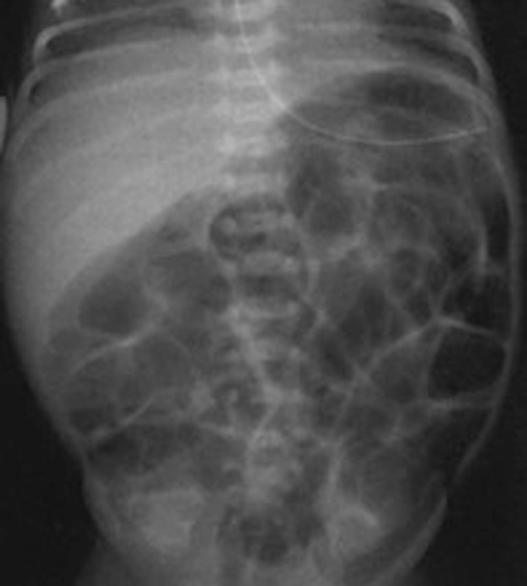
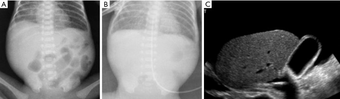
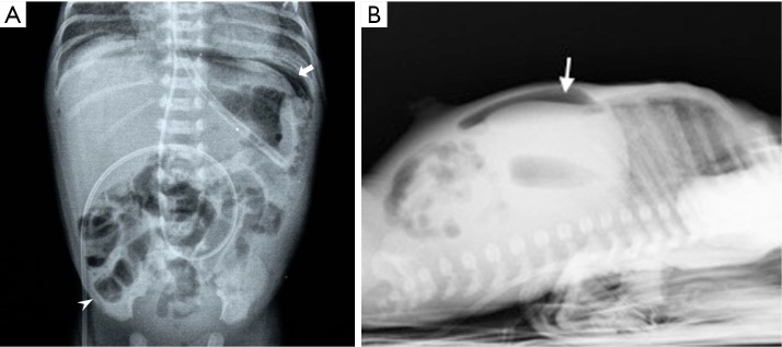
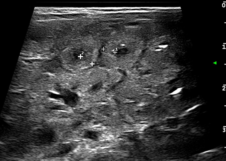
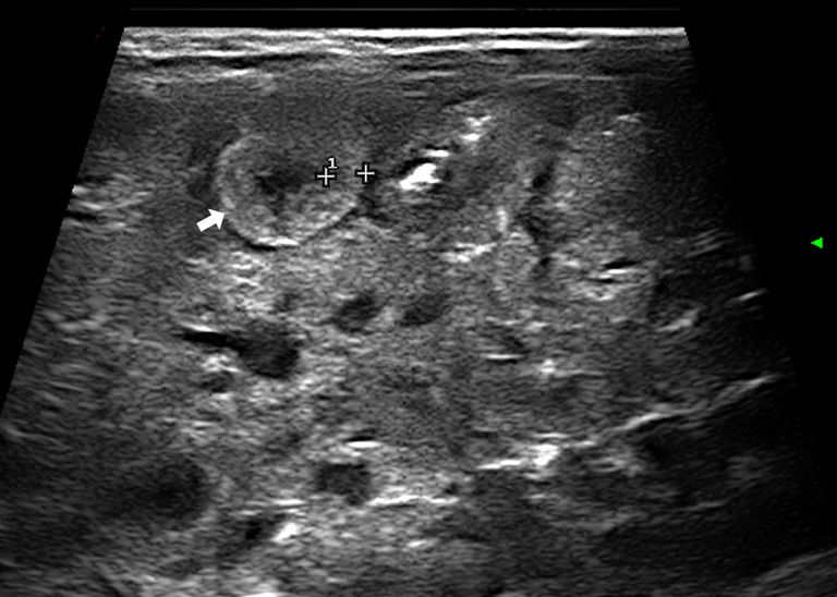
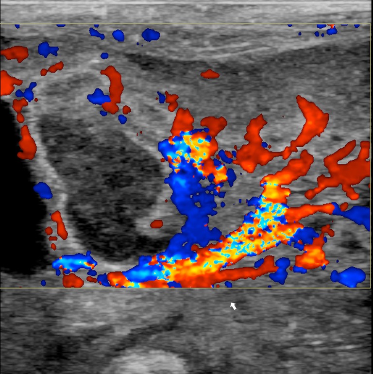
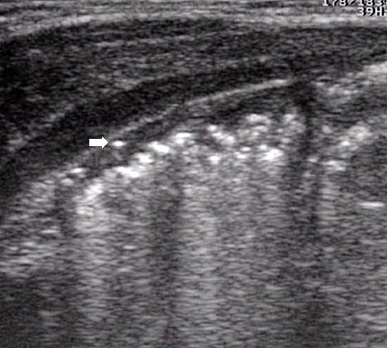
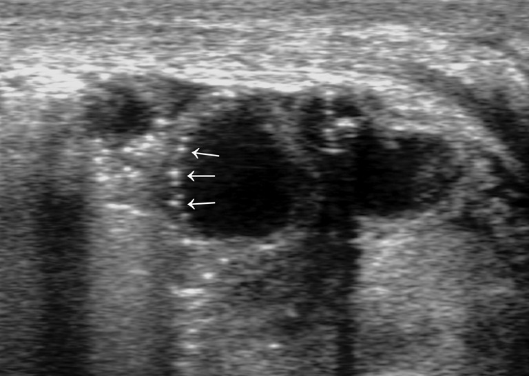
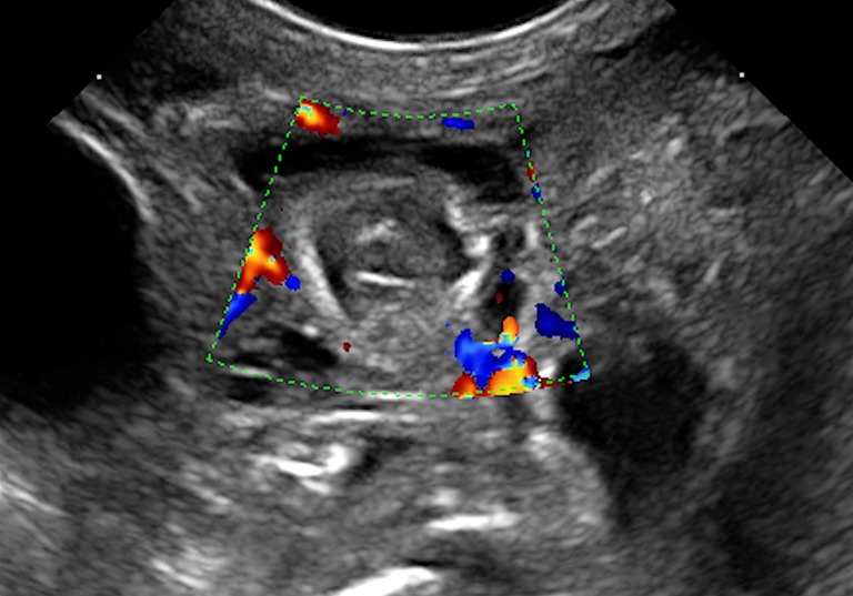
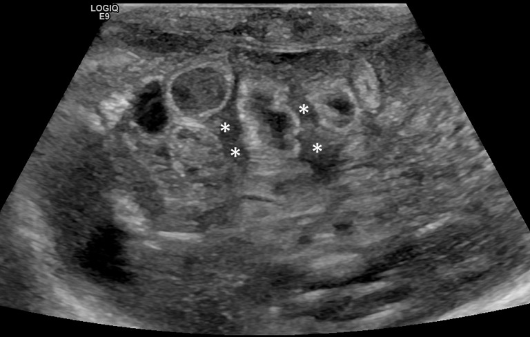
Necrotizing enterocolitis (NEC) is an inflammatory process, characterized by intestinal necrosis of variable extension, leading to perforation, generalized peritonitis and death. The classical pathogenetic theory focuses on mucosal damage related to a stress induced intestinal ischemia leading to mucosal injury and bacterial colonization of the wall. A more recent hypothesis emphasizes the role of immaturity of gastrointestinal and immune system, particularly of the premature, responsible of bowel wall vulnerability and suffering. NEC is the most common gastrointestinal emergency in the newborn, with a higher incidence in the preterm; improvement of neonatal resuscitation techniques enables the survival of premature of very low birth weight (VLBW) with prolongation of hospital stay for perinatal and neonatal care and a higher risk of NEC. Clinical presentation of NEC in newborn ranges from mild forms with moderate gastrointestinal tract disorder and that can heal spontaneously, to very serious forms with fulminant course characterized by perforation, peritonitis, sepsis, disseminated intravascular coagulation (DIC) and shock. Imaging modality in the diagnosis of NEC is historically represented by the plain-film abdominal radiographs which can be performed every 6 hours because of the rapid evolution that may occur in the patient's clinical condition. However ultrasound (US), in recent years, is playing an increasingly important role in the evaluation of early stages of the disease as it provides images in real time of the abdominal structures being able to assess the presence and validity of peristalsis of the bowel loops, detect the thickness of the intestinal wall and the presence of minimal amounts of fluid in the peritoneal cavity. In this paper we review the pathogenesis, clinical presentation and imaging of NEC with a particular attention to the emergent role of US in the diagnosis of the disease.

Keywords: Necrotizing enterocolitis (NEC); bowel disease; newborn; ultrasound (US).


Conflict of interest statement

Conflicts of Interest: The authors have no conflicts of interest to declare.

Figures

Figure 1
Figure 1
Supine plain X-ray of the abdomen: a specific distension of small bowel loops.
Figure 2
Figure 2
Supine plain X-ray showing bowel loops distension with progressive reduction of intestinal air. Ultrasound (US) examination showed an abundant ascites.
Figure 3
Figure 3
Plain X-ray of the abdomen in orthostasis: subdiaphragmatic air (arrow) and intestinal pneumatosis (arrow head).
Figure 4
Figure 4
Bowel wall thickening (>2.6 mm) (caliper).
Figure 5
Figure 5
Disappearance of wall stratification with increased echogenicity (arrow).
Figure 6
Figure 6
Colour Doppler imaging showing increased wall vascularization (arrow) and of mesenterial vessels.
Figure 7
Figure 7
Intestinal pneumatosis (initial phase): presence of hyperechoic spots within the bowel wall (arrow).
Figure 8
Figure 8
Bowel wall pneumatosis (advanced phase): multiple hyperechoic spots within bowel wall. Gas bubbles interest a limited portion of the wall (arrows).
Figure 9
Figure 9
Bowel wall pneumatosis (advanced phase): multiple hyperechoic spots within bowel wall. Gas bubbles interest the entire wall (arrow).
Figure 10
Figure 10
Presence of air microbubbles within the main portal vein (arrow).
Figure 11
Figure 11
Air microbubbles within intrahepatic portal branches as linear hyperechoic spots (arrows).
Figure 12
Figure 12
Bowel wall thinning (arrow). Ultrasound (US) exam shows fine particulate bowel lumen content (*) surrounded by free clear fluid (arrowhead).
Figure 13
Figure 13
Absence of bowel wall vascularization at Colour Doppler examination.
Figure 14
Figure 14
Intraperitoneal free fluid in a newborn with necrotizing enterocolitis (NEC) and perforation: inhomogeneous free fluid between bowel loops with internal echoes and septa (*). In some intestinal loops hyperechoic spots within the wall are present.

References

    1. Lin PW, Stoll BJ. Necrotising enterocolitis. Lancet 2006;368:1271-83. 10.1016/S0140-6736(06)69525-1 - DOI - PubMed
    1. Berman L, Moss RL. Necrotizing enterocolitis: An update. Semin Fetal Neonatal Med 2011;16:145-50. 10.1016/j.siny.2011.02.002 - DOI - PubMed
    1. Dominguez KM, Moss RL. Necrotizing Enterocolitis. Clin Perinatol 2012;39:387-401. 10.1016/j.clp.2012.04.011 - DOI - PubMed
    1. Jona JZ . Advances in neonatal surgery. Pediatr Clin North Am 1998;45:605-17. 10.1016/S0031-3955(05)70031-6 - DOI - PubMed
    1. Kliegman RM, Fanaroff AA. Neonatal necrotizing enterocolitis: a nine-year experience. Am J Dis Child 1981;135:603-7. 10.1001/archpedi.1981.02130310009005 - DOI - PubMed

LinkOut - more resources