Parvovirus Infection Is Associated With Myocarditis and Myocardial Fibrosis in Young Dogs
- PMID: 28812526
- PMCID: PMC10984720
- DOI: 10.1177/0300985817725387
Parvovirus Infection Is Associated With Myocarditis and Myocardial Fibrosis in Young Dogs
Abstract
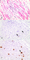
Perinatal parvoviral infection causes necrotizing myocarditis in puppies, which results in acute high mortality or progressive cardiac injury. While widespread vaccination has dramatically curtailed the epidemic of canine parvoviral myocarditis, we hypothesized that canine parvovirus 2 (CPV-2) myocardial infection is an underrecognized cause of myocarditis, cardiac damage, and/or repair by fibrosis in young dogs. In this retrospective study, DNA was extracted from formalin-fixed, paraffin-embedded tissues from 40 cases and 41 control dogs under 2 years of age from 2007 to 2015. Cases had a diagnosis of myocardial necrosis, inflammation, or fibrosis, while age-matched controls lacked myocardial lesions. Conventional polymerase chain reaction (PCR) and sequencing targeting the VP1 to VP2 region detected CPV-2 in 12 of 40 cases (30%; 95% confidence interval [CI], 18%-45%) and 2 of 41 controls (5%; 95% CI, 0.1%-16%). Detection of CPV-2 DNA in the myocardium was significantly associated with myocardial lesions ( P = .003). Reverse transcription quantitative PCR amplifying VP2 identified viral messenger RNA in 12 of 12 PCR-positive cases and 2 of 2 controls. PCR results were confirmed by in situ hybridization, which identified parvoviral DNA in cardiomyocytes and occasionally macrophages of juvenile and young adult dogs (median age 61 days). Myocardial CPV-2 was identified in juveniles with minimal myocarditis and CPV-2 enteritis, which may indicate a longer window of cardiac susceptibility to myocarditis than previously reported. CPV-2 was also detected in dogs with severe myocardial fibrosis with in situ hybridization signal localized to cardiomyocytes, suggesting prior myocardial damage by CPV-2. Despite the frequency of vaccination, these findings suggest that CPV-2 remains an important cause of myocardial damage in dogs.
Keywords: canine parvovirus; cardiomyopathy; dogs; heart; in situ hybridization; myocardial fibrosis; myocarditis.
Conflict of interest statement
Declaration of Conflicting Interests
The author(s) declared no potential conflicts of interest with respect to the research, authorship, and/or publication of this article.
Figures


References
-
- Bastianello SS. Canine parvovirus myocarditis: clinical signs and pathological lesions encountered in natural cases. J S Afr Vet Assoc. 1981;52(2):105–108. - PubMed
-
- Bishop SP. Effect of aortic stenosis on myocardial cell growth, hyperplasia, and ultrastructure in neonatal dogs. Recent Adv Stud Cardiac Struct Metab. 1973;3:637–656. - PubMed
-
- Brinkhof B, Spee B, Rothuizen J, et al. Development and evaluation of canine reference genes for accurate quantification of gene expression. Anal Biochem. 2006;356(1):36–43. - PubMed
MeSH terms
Grants and funding
LinkOut - more resources
Full Text Sources
Other Literature Sources
Medical

