. 2018 Mar;11(2):357-368.
doi: 10.1038/mi.2017.55. Epub 2017 Aug 16.

Bacteroidales recruit IL-6-producing intraepithelial lymphocytes in the colon to promote barrier integrity

Affiliations

Bacteroidales recruit IL-6-producing intraepithelial lymphocytes in the colon to promote barrier integrity

K A Kuhn et al. Mucosal Immunol. 2018 Mar.

Abstract

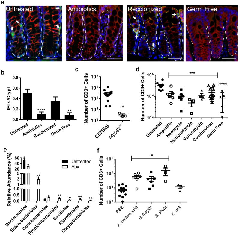
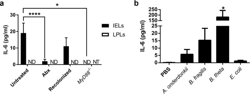
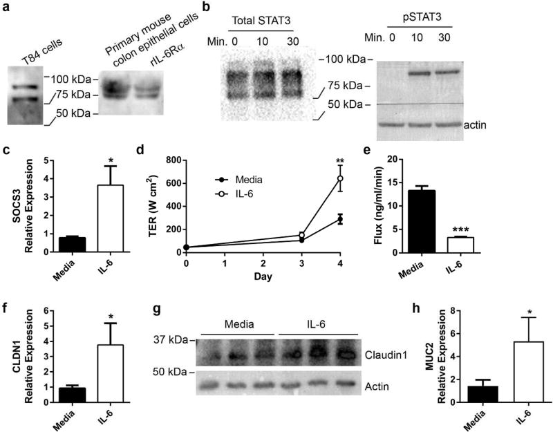
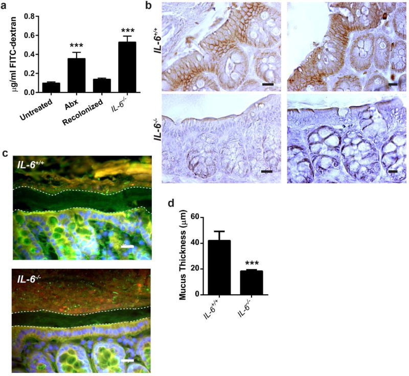
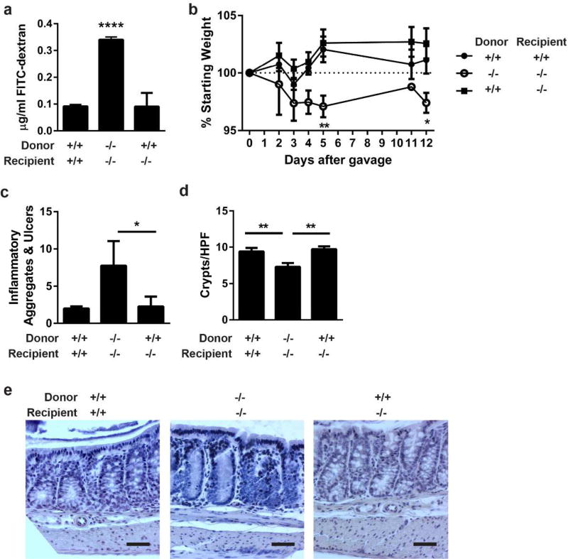
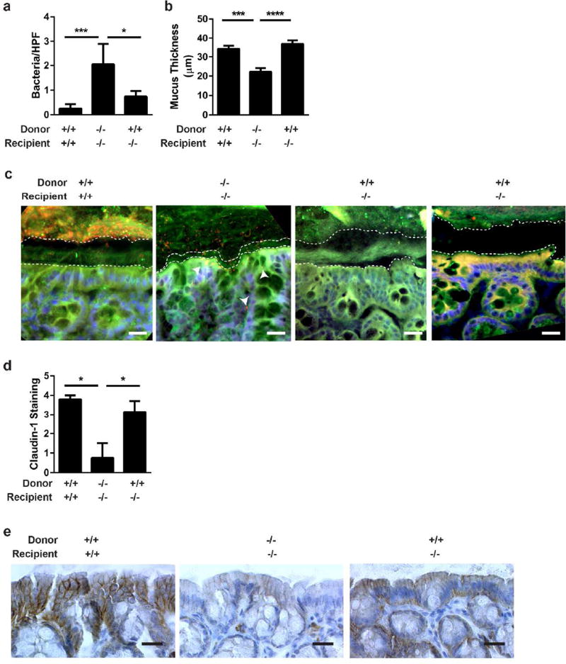
Interactions between the microbiota and distal gut are important for the maintenance of a healthy intestinal barrier; dysbiosis of intestinal microbial communities has emerged as a likely contributor to diseases that arise at the level of the mucosa. Intraepithelial lymphocytes (IELs) are positioned within the epithelial barrier, and in the small intestine they function to maintain epithelial homeostasis. We hypothesized that colon IELs promote epithelial barrier function through the expression of cytokines in response to interactions with commensal bacteria. Profiling of bacterial 16S ribosomal RNA revealed that candidate bacteria in the order Bacteroidales are sufficient to promote IEL presence in the colon that in turn produce interleukin-6 (IL-6) in a MyD88 (myeloid differentiation primary response 88)-dependent manner. IEL-derived IL-6 is functionally important in the maintenance of the epithelial barrier as IL-6-/- mice were noted to have increased paracellular permeability, decreased claudin-1 expression, and a thinner mucus gel layer, all of which were reversed by transfer of IL-6+/+ IELs, leading to protection of mice in response to Citrobacter rodentium infection. Therefore, we conclude that microbiota provide a homeostatic role for epithelial barrier function through regulation of IEL-derived IL-6.


Conflict of interest statement

Disclosure

The authors declare no commercial or financial conflict of interest.

Figures

Figure 1
Figure 1. Bacteria in the class Bacteroides maintain the colonic IEL population
(a–d) Groups of 3–9 C57Bl/6 male and female mice aged 8–12 weeks were untreated, treated with antibiotics for one week, or treated with antibiotics followed by recolonization for one week each. Data are from two independent experiments. (a) Immunofluorescence of methacarn-fixed, paraffin embedded colon tissue from mice was performed and representative images shown at 40X. Bar=20 µm. Dotted lines outline crypts and arrows point to IELs as identified as CD3+ (green) cells in the epithelial layer (β-catenin, red). Nuclei were stained with bis-benzimide (blue). Dashed lines outline crypts while arrows indicate IELs. (b) CD3+ epithelial cells were counted in four well-oriented high-powered fields (HPF) from immunofluorescence staining performed in 6 untreated, 7 antibiotic-treated, 6 recolonized, and 3 germ free mice and shown as the mean number of cells per HPF ± SEM. **, P<0.01 as determined by Kruskall-Wallis analysis with Dunn’s post-test. (c,d) The absolute number of epithelial CD3+ cells harvested from the colons of mice was determined by flow cytometry. Each dot represents an individual mouse and bars are the mean ± SEM. Statistical analysis for (c) was performed using a two-tailed Student’s t-test; **, P<0.01. In (d) statistical analysis was performed using a one-way ANOVA with Dunnett’s multiple comparisons test; ***, P<0.001 and ****, P<0.0001. (e) 16S rRNA sequencing from fecal DNA extracted from 5 untreated and 5 antibiotic-treated mice was performed. Order level differences in relative abundances ± SEM are shown with Wilcoxon rank test performed for statistical analysis. *, P<0.05; **, P<0.01 (f) Germ-free male and female mice aged 8–12 weeks were gavaged with PBS, Alistipes onderdonkii, Bacteroides fragilis, or Bacteroides thetaiotamicron and allowed to colonize for two weeks. Epithelial cells were harvested and CD3+ cells evaluated by flow cytometry. Each dot represents an individual mouse and bars are the mean ± SEM. ***, P<0.001 as determined by one-way ANOVA with Dunnett’s post-test.
Figure 2
Figure 2. IELs utilize bacterial signals for stimulation of IL-6 secretion
IELs were harvested from colons of mice and mitogen-stimulated ex vivo. IL-6 secretion into the supernatant was measured by ELISA, and IL-6 from unstimulated IELs was subtracted from that of the mitogen-stimulated IELs from the same mouse. Data are from groups of 3–9 male and female mice aged 8–12 weeks in two independent experiments and shown as the mean ± SEM. Statistical significance was determined by one-way ANOVA with Dunnett’s test. (a) IL-6 secretion from IELs and LPLs isolated from untreated C57Bl/6 mice as well as antibiotic-treated, recolonized, and MyD88−/− mice. *, P<0.05; ****, P<0.0001. ND = not detected; NT = not tested. (b) IEL secretion of IL-6 from germ-free mice gavaged with PBS or monocolonized with bacteria. *,P<0.05
Figure 3
Figure 3. IL-6 signals in colon epithelia and enhances epithelial barrier function via induction of claudin-1 and mucin-2
(a) IL-6Rα protein in T84 and primary murine epithelial cells was determined by Western blot. (b) T84 colonic epithelial cells were cultured to confluence in the absence or presence of 50 ng/ml recombinant human IL-6. Protein was harvested after 0, 10, and 30 minutes of IL-6 exposure. Western blot confirmed phosphorylation of STAT3 after IL-6 exposure. (c) After 24 hours of IL-6 exposure, RNA from T84 cells was harvested and evaluated by qPCR for SOCS3 expression and normalized to actin. Data are the mean ± SEM fold induction of SOCS3 in IL-6 treated cells compared to untreated cells. An unpaired two-tailed Student’s t-test was used to determine statistical significance. *, P<0.05 (d) T84 cells were cultured on membrane permeable supports in the absence or presence of IL-6. Transepithelial resistance (TER) was recorded daily and shown as the mean ± SEM. An unpaired two-tailed Student’s t-test was performed at each time point to determine statistical significance. **, P<0.01 (e) T84 transwells were evaluated for paracellular flux of FITC-dextran. The rate of flux is shown as the mean ± SEM. An unpaired two-tailed Student’s t-test demonstrated significance. ***, P<0.0001 (f) After 24 hours of IL-6 exposure, RNA from T84 cells was extracted evaluated for CLDN1 expression by qPCR. Data are the mean expression of CLDN1 ± SEM in IL-6 treated cells relative to untreated cells. Statistical analysis using an unpaired two-tailed Student’s t-test revealed significance. *P<0.05 (g) Cellular lysates from unexposed and IL-6 exposed T84 cells at 24 hours were evaluated for claudin-1 protein by Western blot. (h) RNA from T84 cells with and without IL-6 treatment was evaluated for MUC2 expression by qPCR. Data are the mean fold induction of MUC2 ± SEM in treated cells compared to untreated cells. Statistical analysis using an unpaired two-tailed Student’s t-test revealed significance. *P<0.05
Figure 4
Figure 4. Epithelial barrier integrity is impaired in the absence of IL-6
8–12 week old male and female C57Bl/6 and IL-6−/− mice were treated as previously described. Experiments were performed using littermates in groups of 3 mice and repeated twice. (a) Untreated, antibiotic-treated (Abx), recolonized, and IL-6−/− mice were gavaged with 0.6 mg/kg body weight 4 kDa FITC-dextran. After four hours, sera were collected from the mice, the fluorescence at 492 nm measured, and the amount of dextran calculated. Data are the mean concentration of dextran ± SEM. Statistical analyses were performed by one-way ANOVA with Dunnet’s test. ***, P<0.001 (b) Claudin-1 (brown) in vivo was evaluated by immunohistochemistry. Representative photos from 1 of 5 IL-6+/+ and 1 of 4 IL-6−/− mice are shown at 400X. Bar=20 µm. (c) Fluorescent in situ hybridization using a universal bacterial probe (red) was performed on IL-6+/+ and IL-6−/− mice. Nuclei were labeled with bis-benzimide (blue) and the mucus layer labeled with wheat germ agglutinin (green). Representative images are shown at 400X. Bar=20 µm. (d) Measurement of the mucus layer was performed in 3 areas of each of 4–6 high-powered fields (400X) per mouse (6 IL-6+/+ and 6 IL-6−/− mice). Data are the mean ± SEM mucus thickness. An unpaired two-tailed Student’s t-test demonstrated significance. ***, P<0.001
Figure 5
Figure 5. IEL produced IL-6 repairs the epithelial barrier and protects from C. rodentium colitis
(a) IELs from IL-6+/+ or IL-6−/− mice were harvested, magnetically sorted, and transferred into recipient IL-6+/+ or IL-6−/− mice. After one week, intestinal barrier permeability was evaluated by FITC-dextran flux. Two experiments of 2–3 male and female mice per group were performed. Data are the mean serum concentration of dextran ± SEM. ****, P<0.0001 by one-way ANOVA with Tukey’s test. (b) Two days following IEL transfer, 4–7 mice per group were infected with C. rodentium by oral gavage and monitored by daily weights. Mice were euthanized 12 days after infection. Data are the mean percentage of starting weight ± SEM. A two-way repeated-measures ANOVA with Dunnett’s test determined statistical significance. *, P<0.05; **,P<0.01 (c) Methacarn-fixed, paraffin embedded colon tissue from 12-day infected mice in (b) were stained by H&E and evaluated in a blinded fashion for histologic damage as assessed by the number of organized inflammatory aggregates and ulcers along the entire colon. These are shown as the mean ± SEM for each treatment group. *, P<0.05 by Kruskall-Wallis test with Dunn’s multiple comparisons test. (d) Five well-oriented high-powered fields (HPF) per mouse were viewed at 200X and number of crypts counted in each section. Data are the mean crypts/HPF ± SEM. **, P<0.01 by one-way ANOVA with Tukey’s test. (e) Representative histology is shown at 200X. Bar = 50 µm.
Figure 6
Figure 6. Transfer of IL-6+ IELs into IL-6 −/− mice restores the mucus layer and claudin-1 expression
(a) FISH was performed on tissues from Figure 5 and the number of bacteria located within intestinal tissue in each of 20 HPF (at 400X) were counted and shown at the mean bacteria/HPF ± SEM. *, P<0.05; ***,P<0.001 by Kruskall-Wallis test with Dunn’s multiple comparisons test. (b) Mucus thickness in 3 areas of each of 4 HPF per mouse was measured in sections from (a) and displayed as the mean mucus thickness ± SEM. Statistical significance was determined by one-way ANOVA with Tukey’s post-test. ***,P<0.001; ****P<0.0001 (c) Representative FISH is shown at 400X. Bar=20 µm. A universal bacterial probe (red) was used to mark bacteria; nuclei were labeled with bis-benzimide (blue); and the mucus layer labeled with wheat germ agglutinin (green). Dashed white lines outline the epithelial and luminal borders of the inner mucus layer. Arrowheads point to areas of bacterial translocation. (d) Claudin-1 protein expression from the experiment in Figure 5 was evaluated by immunohistochemistry and assessed a numeric score for each mouse based on the level of staining: 0=no staining, 1=faint, 2=mild; 3=moderate; 4=intense. The mean staining intensity per group ± SEM is shown and statistical significance was assessed using a Kruskall-Wallis test with Dunn’s multiple comparisons test. *,P<0.05 (e) Representative claudin-1 immunohistochemistry (brown) is shown at 400X. Bar=20 µm.

References

    1. Lozupone CA, Stombaugh JI, Gordon JI, Jansson JK, Knight R. Diversity, stability and resilience of the human gut microbiota. Nature. 2012;489:220–230. - PMC - PubMed
    1. Hooper LV, Macpherson AJ. Immune adaptations that maintain homeostasis with the intestinal microbiota. Nature reviews. Immunology. 2010;10:159–169. - PubMed
    1. Cheroutre H, Lambolez F, Mucida D. The light and dark sides of intestinal intraepithelial lymphocytes. Nat Rev Immunol. 2011;11:445–456. - PMC - PubMed
    1. Higgins JM, et al. Direct and regulated interaction of integrin alphaEbeta7 with E-cadherin. J Cell Biol. 1998;140:197–210. - PMC - PubMed
    1. Yu Q, et al. MyD88-dependent signaling for IL-15 production plays an important role in maintenance of CD8 alpha TCR alpha beta and TCR gamma delta intestinal intraepithelial lymphocytes. Journal of immunology. 2006;176:6180–6185. - PubMed

Publication types

MeSH terms