Review
. 2018;15(4-5):537-553.
doi: 10.1080/15476286.2017.1356562. Epub 2017 Sep 21.

Celebrating wobble decoding: Half a century and still much is new

Affiliations
Review

Celebrating wobble decoding: Half a century and still much is new

Paul F Agris et al. RNA Biol. 2018.

Abstract

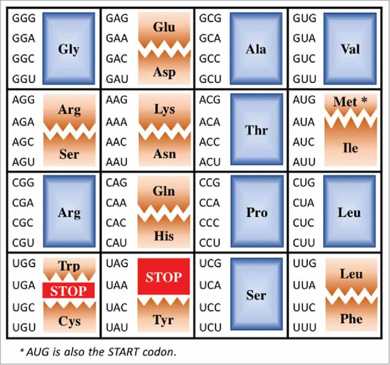
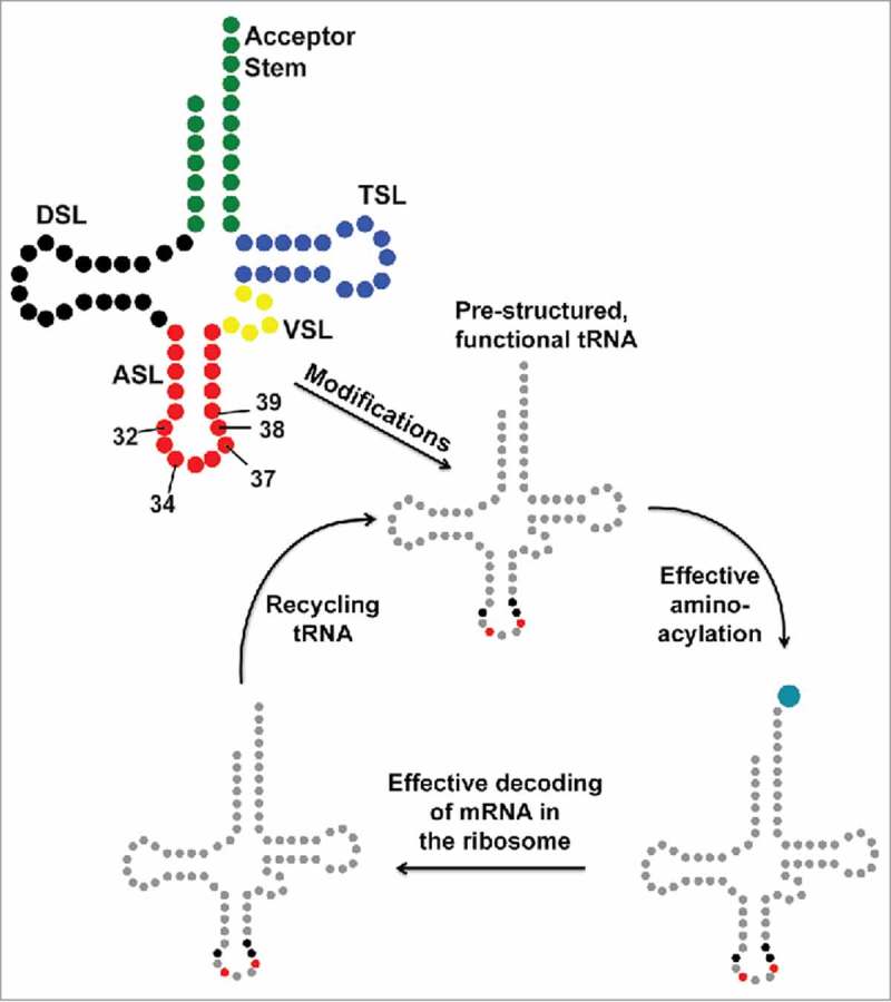
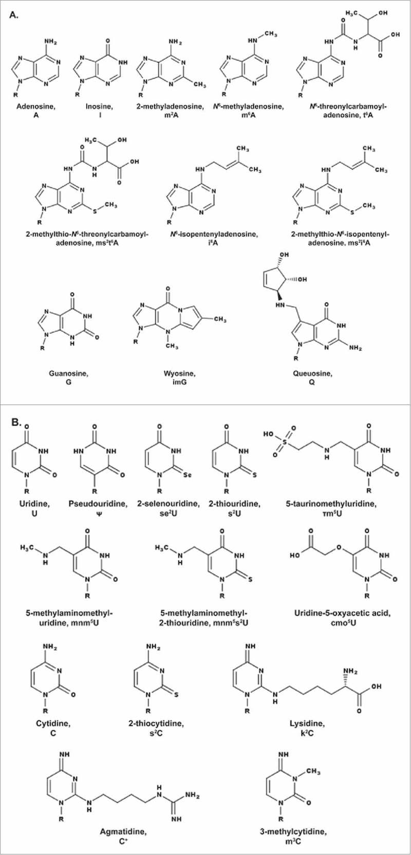
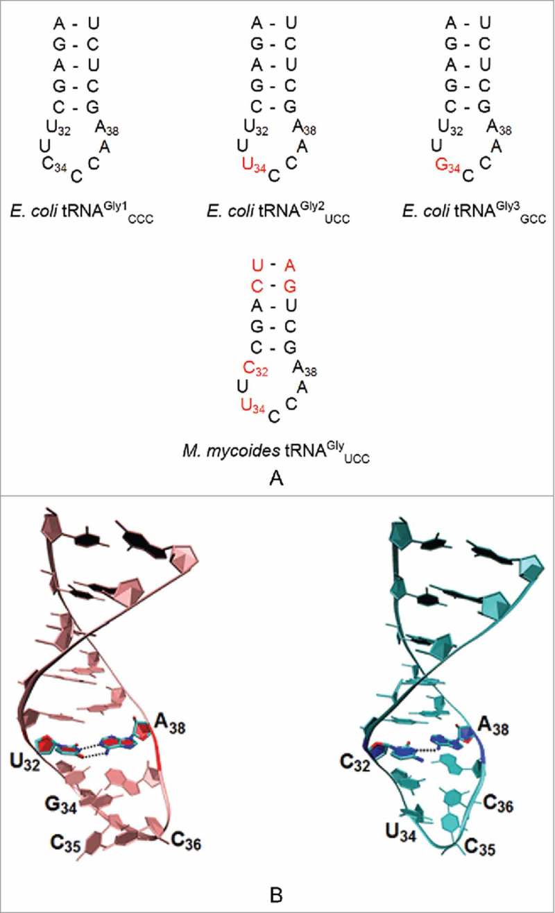
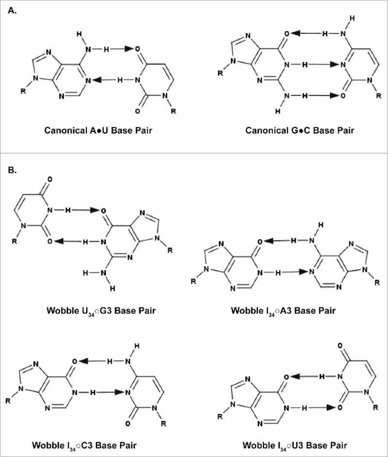
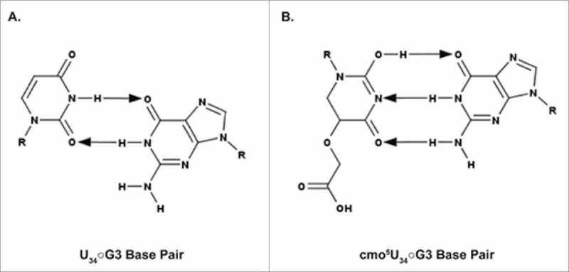
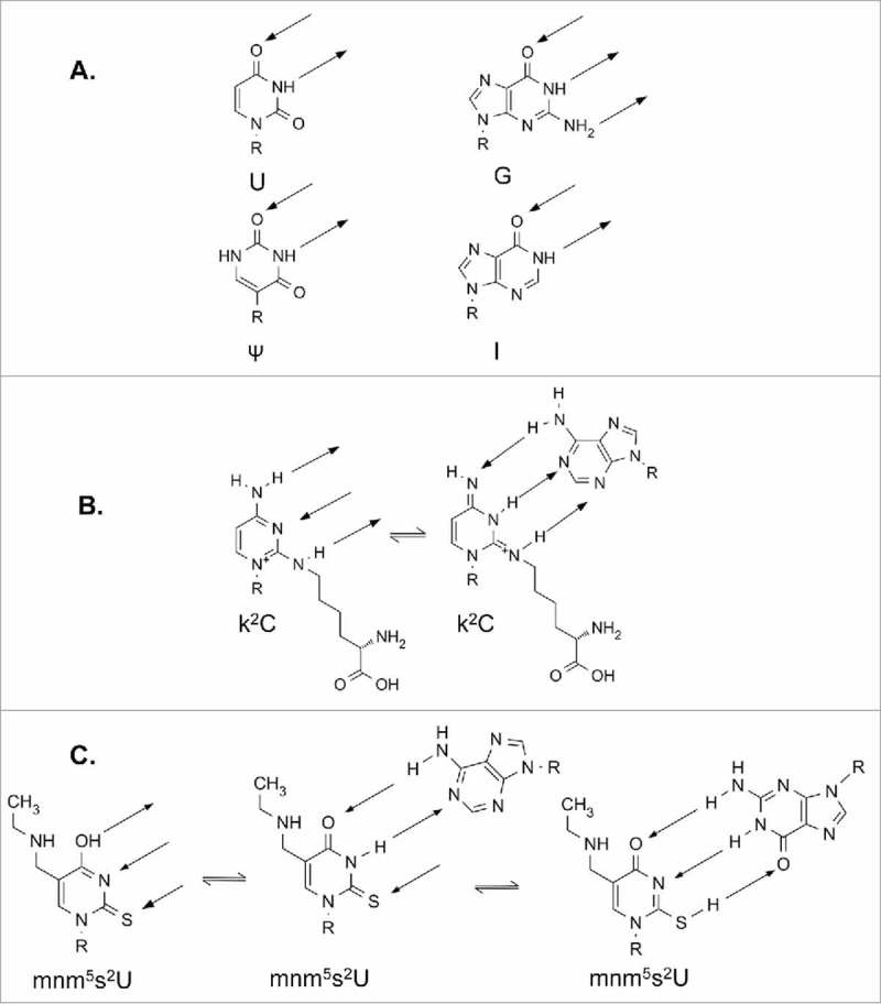
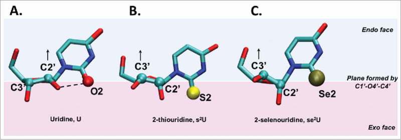
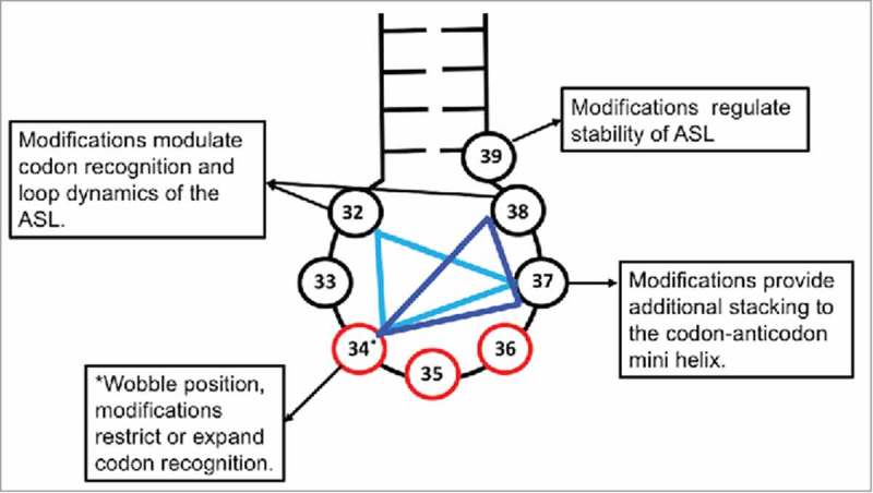
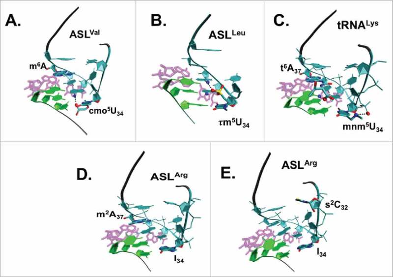
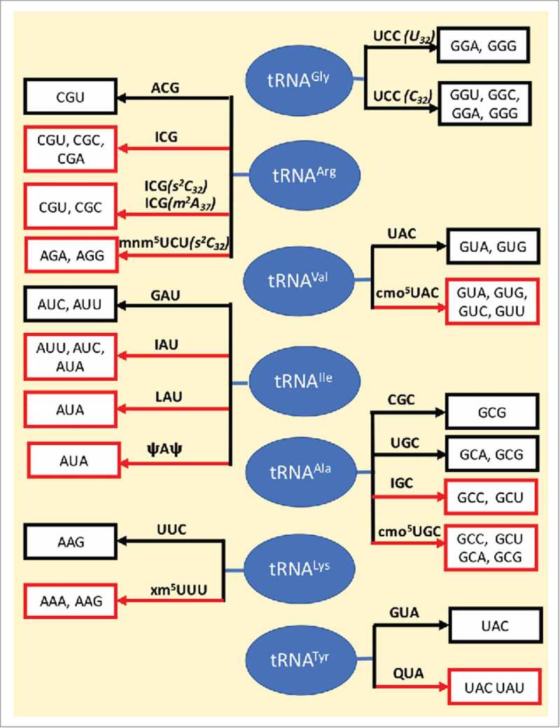
A simple post-transcriptional modification of tRNA, deamination of adenosine to inosine at the first, or wobble, position of the anticodon, inspired Francis Crick's Wobble Hypothesis 50 years ago. Many more naturally-occurring modifications have been elucidated and continue to be discovered. The post-transcriptional modifications of tRNA's anticodon domain are the most diverse and chemically complex of any RNA modifications. Their contribution with regards to chemistry, structure and dynamics reveal individual and combined effects on tRNA function in recognition of cognate and wobble codons. As forecast by the Modified Wobble Hypothesis 25 years ago, some individual modifications at tRNA's wobble position have evolved to restrict codon recognition whereas others expand the tRNA's ability to read as many as four synonymous codons. Here, we review tRNA wobble codon recognition using specific examples of simple and complex modification chemistries that alter tRNA function. Understanding natural modifications has inspired evolutionary insights and possible innovation in protein synthesis.

Keywords: Modified Wobble Hypothesis; Wobble Hypothesis; cognate and wobble codon recognition; modified nucleosides; nucleoside tautomers; tRNA; translation; wobble decoding; wobble nucleoside.


Figures

Figure 1.
Figure 1.
The Universal Genetic Code. The Universal Genetic Code is shown in an atypical array to highlight those codons and their decoding by tRNAs discussed here. Fully degenerate codon boxes are shown in blue, split codon boxes in brown and stop codons in red.
Figure 2.
Figure 2.
The tRNA journey. The secondary structure of tRNA with its constituent domains marked in different colors (top left): Acceptor Stem (green); Dihydrouridine Stem and Loop, DSL (black); Anticodon Stem and Loop, ASL (red); Variable Stem and Loop, VSL (yellow); Thymidine Stem and Loop, TSL (blue). tRNA transcripts are processed by sizing and modification, some are spliced, before functioning in translation. Modification of tRNAs, particularly the anticodon stem and loop (ASL) domain at positions 32, 34, 37, 38 and 39, is an important step toward achieving functional chemistry and architecture. The wobble nucleoside, first of the anticodon, is position 34. Red and black highlights of mature tRNA after modification indicate the locations in the ASL where it is heavily modified.
Figure 3.
Figure 3.
Modifications present in tRNA's anticodon stem and loop domain (ASL) featured in the text and displayed in their neutral state. A. Modified purines, adenosine (A) and guanosine (G). B. Modified pyrimidines, uridine (U) and cytidine (C). R = ribose.
Figure 4.
Figure 4.
Sequences of unmodified ASL of tRNAGly. A. ASLs of E. coli tRNAGly1 CCC, tRNAGly2 UCC, and tRNAGly3 GCC indicating the changes in nucleoside position 32 and 34 (marked in red) from the tRNAGly1 sequence. All 3 E. coli tRNAGly have the same nucleoside at position 32 (U32). The Mycoplasma mycoides tRNAGly UCC differs from the E. coli tRNAGly as a result of a cytidine at position 32 as well as the presence of inverted base pairs in the anticodon stem at positions 27•43 and 28•42. B. Three Dimensional Structure of the tRNAGly GCC and tRNAGly UCC ASLs of B. subtilis. The interactions between the U32•A38 and C32○A38 nucleosides (PDB ID: 2LBJ and 2LBL) are shown. Both ASLs lack the sharp U-turn characteristic of ASLs of other tRNAs. Nucleosides of the anticodon are labeled 34 to 36.
Figure 5.
Figure 5.
Canonical and wobble base pairing of tRNA to mRNA. A. Canonical A•U and G•C base pairs. B. Wobble U34○G3, I34○A3, I34○C3, and I34○U3 base pairs. G34○U3 pairings are virtually nonexistent; therefore, the pairing is not shown. The arrows point away from the hydrogen bond donor and toward the hydrogen bond acceptor.
Figure 6.
Figure 6.
Watson-Crick geometry of the cmo5U34○G3 base pair. A. The predicted geometry of the unmodified U34○G3 base pair containing two hydrogen bonds. B. The observed geometry of cmo5U34○G3 base pair containing three hydrogen bonds. For the cmo5U34○G3 to resemble a C•G base pair, the cmo5 modification is proposed to facilitate formation of the enol tautomer. The arrows point away from the hydrogen bond donor and toward the hydrogen bond acceptor.
Figure 7.
Figure 7.
Modifications influence protein and codon recognition of tRNA through altered hydrogen bonding. R represents the ribose sugar. Hydrogen bonding is indicated by arrows. A. Nucleosides commonly found at position 34 of tRNAIle. All 4 bases are capable of recognition by isoleucyl-tRNA synthetase, IleRS. B. Two possible tautomers of lysidine. Structure on the right can be recognized by IleRS, and could putatively recognize A as shown. C. Possible tautomers of mnm5s2U34 of E. coli tRNALys UUU. Likely interactions with A and G are shown.
Figure 8.
Figure 8.
Sulfur and selenium atomic radii affect sugar pucker when modifying the C2 position of uridine. A. Uridine, B. 2-thiouridine and C. 2-selenouridine nucleosides are shown. All 3 nucleosides are depicted in their anti conformation. The plane defined by atoms C1′-O4′-C4′ of the sugar separates the endo face (the side in which C5′ projects from the plane) from the exo face. In the case of uridine, the sugar pucker is C2′ endo as indicated by atom C2′ in the endo face (above the C1′-O4′-C4′ plane), while in 2-thiouridine and 2-selenouridine, the relative increase in atomic radius of substituent atom X at position C2 and the weakening of the hydrogen bonding interaction between 2′OH and X (where X – O2, S2 or Se2) shifts the equilibrium toward a C3′ endo conformation.
Figure 9.
Figure 9.
ASL modification triangle. A combination of 3 positions in the ASL (nucleotides 32 through 38) of tRNAs are commonly modified to expand or restrict codon recognition. Position 32, 34 and 37 (light blue) or position 34, 37 and 38 (dark blue) form vertices of the triangle in which modifications work in a co-surgical manner to achieve desired ribosomal binding affinity of the tRNA to different codons. The anticodon is represented in red and the nucleosides in the ASL in black.
Figure 10.
Figure 10.
Ribosome-bound structures of the ASLs of 5 tRNA species with modifications. tRNA modified nucleosides at positions 32, 34 and 37 are influential in creating the architecture of the anticodon domain accepted into the ribosome's A-site for cognate and wobble codon binding. The codon on the mRNA is shown in green, the ASL in cyan with the modifications labeled and the rRNA interactions shown in magenta. Possible interactions of the modified groups are represented using a dashed line. A. In ASLVal UAC bound to codon GUU, (PDB ID: 2UUB), the carboxyl oxygen of cmo5U34 is within hydrogen bonding distance of the amine group on the neighboring A35. B. In ASLLeu UAA bound to codon UUG (PDB ID: 2VQF), two of the three oxygens bonded to the sulfur atom in the τm5U34 can form hydrogen bonding interactions with A35 and A36. C. In ASLLys UUU bound to codon AAA (PDB ID: 1XMQ), there are a salt-bridge between the 5-methylaminomethyl group on U34 and its phosphate group, an enhanced stacking interaction provided by t6A37, and a possible hydrogen bond between the threonyl group of t6A37 and A1 on the mRNA. D. ASLArg2 ICG bound to the codon CGC. The ASL has the two modifications I34, m2A37. E. ASLArg1 ICG bound to the codon CGC. The ASL has the modifications s2C32, I34. When either of the modifications m2A37 or s2C32 are present, then the tRNA is unable to recognize the rare CGA codon.39
Figure 11.
Figure 11.
Wobble map. Anticodon domain modified nucleosides play an important role in the wobbling of tRNA's codon recognition, some expanding codon recognition to synonymous codons while others restrict recognition. The different cases of wobbling in the presence and absence of modifications as discussed in this paper, are shown. The black arrow represents no modifications, whereas a red arrow indicates the presence of one or more modifications. The tRNAs are shown in blue, the anticodons are indicated on the arrow and codons decoded by the tRNA bearing the anticodon are enclosed in white boxes.

References

    1. Crick FH. Codon—anticodon pairing: The wobble hypothesis. J Mol Biol. 1966; 19:548–55. doi:10.1016/S0022-2836(66)80022-0. - DOI - PubMed
    1. Holley RW, Apgar J, Everett GA, Madison JT, Marquisee M, Merrill SH, Penswick JR, Zamir A. Structure of a ribonucleic acid. Science. 1965;147:1462–5. doi:10.1126/science.147.3664.1462. - DOI - PubMed
    1. Hall RH. A general procedure for the isolation of “minor” nucleosides from ribonucleic acid hydrolysates. Biochemistry. 1965;4:661–70. doi:10.1021/bi00880a008. - DOI - PubMed
    1. Crick FH. The genetic code—yesterday, today, and tomorrow. Cold Spring Harb Symp Quant Biol. 1966;31:1–9. doi:10.1101/SQB.1966.031.01.007. - DOI - PubMed
    1. Agris PF. Wobble position modified nucleosides evolved to select transfer RNA codon recognition: A modified-wobble hypothesis. Biochimie. 1991;73:1345–9. doi:10.1016/0300-9084(91)90163-U. - DOI - PubMed

Publication types

MeSH terms

LinkOut - more resources