Review
. 2017 Nov/Dec;9(8):1231-1252.
doi: 10.1080/19420862.2017.1367074. Epub 2017 Aug 16.

IgG cooperativity - Is there allostery? Implications for antibody functions and therapeutic antibody development

Affiliations
Review

IgG cooperativity - Is there allostery? Implications for antibody functions and therapeutic antibody development

Danlin Yang et al. MAbs. 2017 Nov/Dec.

Abstract

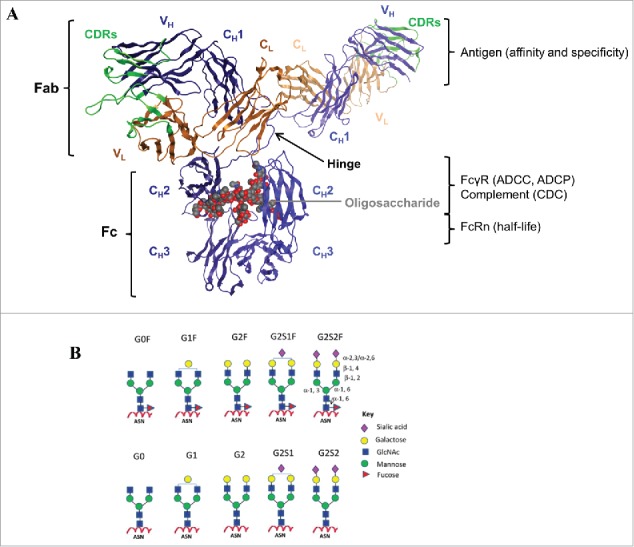
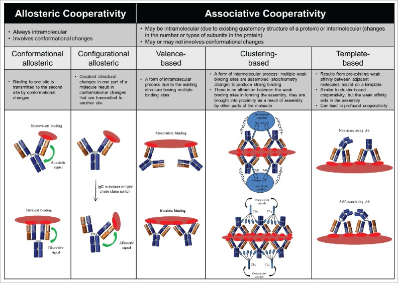
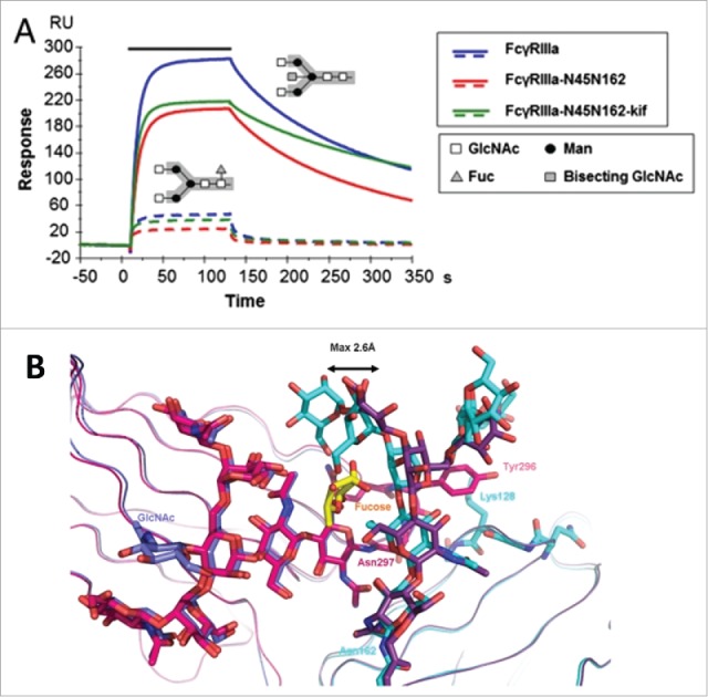
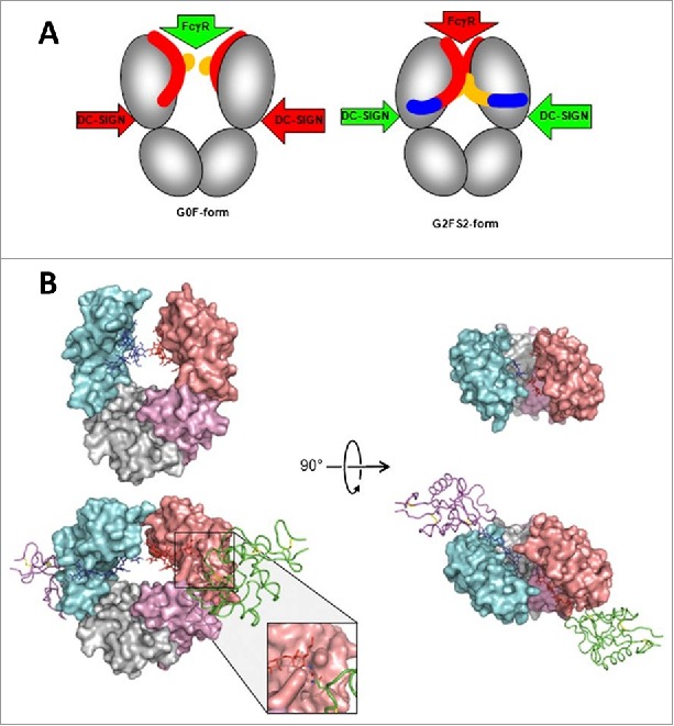
A central dogma in immunology is that an antibody's in vivo functionality is mediated by 2 independent events: antigen binding by the variable (V) region, followed by effector activation by the constant (C) region. However, this view has recently been challenged by reports suggesting allostery exists between the 2 regions, triggered by conformational changes or configurational differences. The possibility of allosteric signals propagating through the IgG domains complicates our understanding of the antibody structure-function relationship, and challenges the current subclass selection process in therapeutic antibody design. Here we review the types of cooperativity in IgG molecules by examining evidence for and against allosteric cooperativity in both Fab and Fc domains and the characteristics of associative cooperativity in effector system activation. We investigate the origin and the mechanism of allostery with an emphasis on the C-region-mediated effects on both V and C region interactions, and discuss its implications in biological functions. While available research does not support the existence of antigen-induced conformational allosteric cooperativity in IgGs, there is substantial evidence for configurational allostery due to glycosylation and sequence variations.

Keywords: IgG allostery; IgG subclass selection; antibody discovery; cooperativity; glycosylation; intermolecular interaction; intramolecular interaction; molecular engineering; structure and function.


Figures

Figure 1.
Figure 1.
IgG structure and function. (A) The crystal structure of a human IgG1 molecule is used to illustrate domain assignments (PDB ID: 1HZH). An individual IgG is composed of 2 identical heavy chains (blue) and 2 identical light chains (orange), linked together by inter-chain disulfide bonds. Each heavy chain consists of a variable (VH) domain and 3 constant (CH1, CH2, and CH3) domains, while the light chain is composed of a variable (VL) and a constant (CL) domain. The light chain pairs with the VH and CH1 domains to form the Fab, which interacts to form the antigen-binding region, also known as the complementarity-determining region (CDR, green). The CH2 and CH3 domains dimerize to form the Fc, which is connected to the upper Fab region via a flexible hinge containing several disulfide bridges that covalently link the CH1 and CH2 chains together. The interactions between the Fc and the Fc-gamma receptors (FcγR) expressed by effector cells or the complement component 1q (C1q), are vital to the clearance of target antigens through antibody-dependent cell-mediated cytotoxicity (ADCC) and phagocytosis (ADCP), or complement-dependent cytotoxicity (CDC), respectively. The highly conserved N-linked glycosylation site (gray) located in the CH2 domains is responsible for the overall structural integrity of the IgG molecule to mediate effector functions. In addition, the Fc can bind the neonatal Fc receptor (FcRn) in a strictly pH-dependent manner, an interaction that contributes to the long serum half-lives of human IgGs. (B) Schematic diagrams of N-linked glycoforms commonly found in human IgGs or industrial mAbs. The N-linked glycoforms attached to asparagine (Asn) 297 are categorized into 2 groups: fucosylated (top panels) and non-fucosylated (bottom panels). A core heptasaccharide structure (G0) is composed of 2 N-acetylglucosamine (GlcNAc), 3 mannose, and 2 GlcNAc residues that are β-1,2 linked to the mannose from the α-1,6 and α-1,3 arms, forming 2 antennae. In addition to fucose, galactose, bisecting GlcNAc, and sialic acid may be added to the core. Figure reproduced from with permission from Elsevier.
Figure 2.
Figure 2.
Classification of the potential forms of cooperativity in IgG and their nomenclature. Please refer to the Appendix I for additional information.
Figure 3.
Figure 3.
Fucosylation-induced allostery on FcγR binding. (A) Comparison of the binding interactions between FcγRIIIA and human IgG1 glycovariants. Overlay of SPR sensorgrams for binding of 125 nM FcγRIIIA glycovariants to fucosylated (dotted line) and afucosylated (continuous line) IgG1s. The association phase is indicated by a solid bar above the curves. The afucosylated IgG significantly enhanced binding to all FcγRIIIA glycovariants with up to 100-fold increase in affinity as compared with the fucosylated version. The N-linked glycosylation is shown in the insert containing the core pentasaccharide (gray box) and the additional carbohydrate residues (legend box). (B) Overlay of the crystal interaction interface between glycosylated FcγRIIIA and Fc glycovariants. Chain A of the afucosylated (blue) bound to FcγRIIIA (cyan) and of the fucosylated (magenta) Fc bound to FcγRIIIA (dark violet) with core fucose (yellow). The oligosaccharide at Asn 162 is displaced by a maximum distance of 2.6 Å in comparison to its position in the structure with a fucosylated Fc. Figure reproduced from an open access article from.
Figure 4.
Figure 4.
A proposed allosteric model for Fc effector function regulation through sialylation. (A) Schematic of the blockade of FcγR binding as a result of sialylation-induced conformational changes within the Fc. The G0F-Fc maintains an “open” conformation (left) that allows FcγR binding, whereas the fully sialylated G2FS2 (blue) interacts with the CH2 domain to induce a “closed” conformation that prevents FcγR binding while revealing the binding site for DC-SIGN. DC-SIGN is an alternative cellular receptor responsible for anti-inflammatory responses (please refer to the article for details). (B) The “open” crystal front (upper left) and top (upper right) view of the G0F-Fc and the “closed” crystal front (lower left) and top (lower right) view of the G2FS2-Fc with DC-SIGN bound. Figure reproduced from with permission from PNAS.
Figure 5.
Figure 5.
Proposed associative models of FcγR crosslinking and activation. (A) The simple avidity model and (B) the ordered receptor aggregation model by Radaev and Sun. Figure reproduced from with permission from Elsevier. The IgG dislocation models proposed by Woof and Burton: (C) Fc array formation from adjacent antigen-bound IgGs facilitates FcγR binding and (D) FcγR binding to distant antigen-bound IgGs coupled with membrane rearrangement facilitates Fc array formation. Figure reproduced from with permission from Macmillan Publishers Ltd.
Figure 6.
Figure 6.
Proposed associative models of IgG hexamerization and complement activation. (A) Structure-function relationship of a triple mutant IgG1 mAb RGY (E345R/E430G/S440Y) in solution: 1) enhanced CDC activity relative to wild-type IgG1–005 and IgG1-E345R; 2) an overview electron tomography (ET) image showing a monomer (small circle) and a hexamer (large circle); 3) a representative hexamer with colored Fab pairs; and 4) ET average of 200 subtomograms at a resolution of 2.9 nm. Figure reproduced from with permission from AAAS. (B) Native mass spectrometry analysis of reconstructed C1, C1:IgG, and C1:IgG:Ag complexes. The assembly C1q:C1r:C1s stoichiometry of 1:2:2 is consistent with the reported composition of natural C1. The C1 exhibits the same IgG binding stoichiometry as C1q. In the presence of excess soluble antigen, the assembly C1:IgG:Ag stoichiometry of 1:6:12 is the predominant species. (C) Model summarizing the molecular determinants for IgG-mediated activation of the classical component pathway: 1) availability of antigen and epitope distribution; 2) ability of antigen to cluster IgG at the cell surface or in solution; 3) Fc-Fc associative cooperativity required for hexamerization; 4) avidity binding sites for hexavalent C1q; 5) composition of Fc oligosaccharide; 6) Fab-Fab intermolecular cooperativity; and 7) antigen-induced conformational allostery to affect downstream Fc-mediated complement activation. Figures reproduced from with permission from Elsevier.
Figure 7.
Figure 7.
Interchain disulfide linkage characteristics and structural isoforms of human IgG subclasses. (A) Schematic comparison of disulfide linkages and hinge amino acid sequences between the subclasses. The core hinge region sequences are displayed under each schematic. (B) Structural isoforms of IgG2 resulting from inter-chain disulfide shuffling: IgG2-A is the known classical form, IgG2-B is created by a symmetric disulfide linkage of both Fab regions to the hinge, and IgG2-A/B is an intermediate form with an asymmetric disulfide linkage of one Fab arm to the hinge. (C) Formation of a bispecific monovalent IgG4 molecule resulting from Fab arm exchange between 2 different monospecific bivalent IgG4 molecules. The non-covalently linked half molecule is created by the formation of intra-chain disulfide bonds as depicted in the insert. The C regions are shown in solid colors and the V regions are patterned.
Figure 8.
Figure 8.
Immunofluorescence patterns of V-region identical chimeric mAbs binding to encapsulated Cryptococcus neoformans cells. Differences in the fluorescence pattern and intensity throughout the capsule are observed: IgG1, IgG2, IgG4, and IgA1 produced an annular pattern, whereas IgG3 and IgM revealed a punctate pattern. IgG4 gave a thick annular pattern that is different from the other subclasses. Figure reproduced from with permission from Copyright 2002. The American Association of Immunologists, Inc.
Figure 9.
Figure 9.
In vivo protective efficacy and capsule reactivity of V-region identical murine IgG subclass mAbs against Bacillus anthracis. (A) Overall survival of mAb-treated mice after receiving lethal dose of Bacillus anthracis spores. Only IgG3 mAbs were protective in a dose-dependent manner; treatment with IgG1, IgG2a, or IgG2b mAbs did not significantly increase the overall survival percentage at any given dose. (B) Incubation of Bacillus anthracis cells with each mAb variant and evaluation by differential interference contrast microscopy. The IgG3 mAbs produced dual-capsule binding reactions at both the outer edge (red arrow) and inner layer near the cell wall (blue arrow); the other subclass variants produced a “puffy” type of reaction where no reactivity to either region was observed. Figure reproduced from an open access article from.
Figure 10.
Figure 10.
SPR sensorgrams of V-region identical (A) intact human IgG subclass mAbs and (B) respective F(ab’) domains binding to mucin immobilized onto the sensor surface. Each set was analyzed using the same sample concentration. IgG1 and IgG2 exhibited faster dissociation than IgG3 and IgG4. IgG4 achieved the highest maximal binding response at the end of the association phase. In contrast, insignificant binding pattern difference was observed between the F(ab’) fragments. IgG4 F(ab’) showeda slightly higher binding response than the others. Figure reproduced from with permission from Wiley.
Figure 11.
Figure 11.
Circular dichroism analysis of V-region identical murine IgG subclass mAbs (A) with and (B) without bound antigen. The antigen-bound subclass pairs IgG1/IgG2b and IgG2a/IgG3 shared similar patterns of changes in secondary structure with difference in magnitude, whereas the secondary structure content was similar between all the subclasses in the absence of antigen. H(d) represents α helix (disordered) secondary structure; S(r) represents β sheet (regular) secondary structure; S(d) represents β sheet (disordered) secondary structure. Figure reproduced from with permission from Elsevier.

References

    1. Chan AC, Carter PJ. Therapeutic antibodies for autoimmunity and inflammation. Nat Rev Immunol. 2010;10:301-16. doi:10.1038/nri2761. PMID:20414204 - DOI - PubMed
    1. Sliwkowski MX, Mellman I. Antibody therapeutics in cancer. Science. 2013;341:1192-8. doi:10.1126/science.1241145. PMID:24031011 - DOI - PubMed
    1. Sharma P, Allison JP. The future of immune checkpoint therapy. Science. 2015;348:56-61. doi:10.1126/science.aaa8172. PMID:25838373 - DOI - PubMed
    1. Melis JPM, Strumane K, Ruuls SR, Beurskens FJ, Schuurman J, Parren PWHI. Complement in therapy and disease: Regulating the complement system with antibody-based therapeutics. Mol Immunol. 2015;67:117-30. doi:10.1016/j.molimm.2015.01.028. PMID:25697848 - DOI - PubMed
    1. Ecker DM, Jones SD, Levine HL. The therapeutic monoclonal antibody market. mAbs. 2015;7:9-14. doi:10.4161/19420862.2015.989042. PMID:25529996 - DOI - PMC - PubMed

MeSH terms