Review
. 2017 Aug 16;17(8):1887.
doi: 10.3390/s17081887.

Recent Advances in Electrospun Nanofiber Interfaces for Biosensing Devices

Affiliations
Review

Recent Advances in Electrospun Nanofiber Interfaces for Biosensing Devices

Eleni Sapountzi et al. Sensors (Basel). .

Abstract

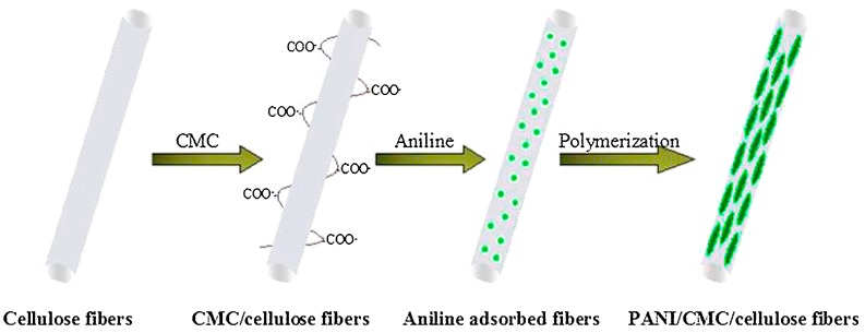
Electrospinning has emerged as a very powerful method combining efficiency, versatility and low cost to elaborate scalable ordered and complex nanofibrous assemblies from a rich variety of polymers. Electrospun nanofibers have demonstrated high potential for a wide spectrum of applications, including drug delivery, tissue engineering, energy conversion and storage, or physical and chemical sensors. The number of works related to biosensing devices integrating electrospun nanofibers has also increased substantially over the last decade. This review provides an overview of the current research activities and new trends in the field. Retaining the bioreceptor functionality is one of the main challenges associated with the production of nanofiber-based biosensing interfaces. The bioreceptors can be immobilized using various strategies, depending on the physical and chemical characteristics of both bioreceptors and nanofiber scaffolds, and on their interfacial interactions. The production of nanobiocomposites constituted by carbon, metal oxide or polymer electrospun nanofibers integrating bioreceptors and conductive nanomaterials (e.g., carbon nanotubes, metal nanoparticles) has been one of the major trends in the last few years. The use of electrospun nanofibers in ELISA-type bioassays, lab-on-a-chip and paper-based point-of-care devices is also highly promising. After a short and general description of electrospinning process, the different strategies to produce electrospun nanofiber biosensing interfaces are discussed.

Keywords: bioreceptor immobilization; biosensing devices; carbon nanofibers; carbon nanotubes; electrospinning; metal nanoparticles; metal oxide nanofibers; polymer nanofibers.


Conflict of interest statement

The authors declare no conflict of interest.

Figures

Figure 1
Figure 1
Typical horizontal (a) and vertical (b) electrospinning set-ups. They are represented with a static collector plate but other configurations exist. Reprinted with permission from [16]. Copyright 2016 Elsevier.
Figure 2
Figure 2
Coaxial (a) and triaxial (b) electrospinning set-ups. Reprinted with permission from [16]. Copyright 2016 Elsevier.
Figure 3
Figure 3
Principle of the electrospun NFs-based immunosensor for amperometric detection α-Fetoprotein proposed by Li et al. Reprinted with permission from [66]. Copyright 2015 American Chemical Society.
Figure 4
Figure 4
(a) TEM, (b) HRTEM, and (c) STEM images of IrOx nanofibers after annealing at 500 °C. The inset of panel (a) is the enlarged TEM image and inset of panel (b) is the SAED pattern. Panels (d,e) are the corresponding EDX mapping of Ir and O elements. Reprint with permission from [66]. Copyright 2015 American Chemical Society.
Figure 5
Figure 5
SEM images of (a) CENFs and (b) Cu/CENFs. Reprint with permission from [42].
Figure 6
Figure 6
Fabrication process of the (a) AgNPs embedded in the PVA water-stable nanofibers and (b) AgNPs immobilized on the functionalized PVA/PEI water-stable nanofibers Reprint with permission from [53]. Copyright 2013 Elsevier.
Figure 7
Figure 7
Preparation of PANI/CMC/cellulose nanofibers. Reprinted with permission from [57]. Copyright 2015 Elsevier.
Figure 8
Figure 8
TEM images of PANI/CMC/cellulose nanofibers at different magnifications. Reprinted with permission from [57]. Copyright 2015 Elsevier.
Figure 9
Figure 9
TEM images of (a) pure PVA-SbQ NFs, (b) PVA-SbQ/MWCNTs (1 wt %) NFs, (c) PVA-SbQ/MWCNTs (5 wt %) NFs, (d) PVA-SbQ/MWCNTs (10 wt %). Reprinted with permission from [82]. Copyright 2015 The Electrochemical Society
Figure 10
Figure 10
SEM images of water-stable electrospun PVA/PEI NFs before immersion in the Au NPs solution (a), after immersion in colloidal Au NPs solutions of pH 7.0 (b), pH 6.0 (c), pH 5.0 (d). Reprinted with permission from [83]. Copyright 2016 Elsevier.

Similar articles

Cited by

References

    1. Karimian N., Moretto L.M., Ugo P. Nanobiosensing with arrays and ensembles of nanoelectrodes. Sensors. 2017;17:65. doi: 10.3390/s17010065. - DOI - PMC - PubMed
    1. Lopez G.A., Estevez M.-C., Solera M., Lechuga L.M. Recent advances in nanoplasmonic biosensors: Applications and lab-on-a-chip integration. Nanophotonics. 2017;6:123–136. doi: 10.1515/nanoph-2016-0101. - DOI
    1. Kim J., Kumar R., Bandodkar A.J., Wang J. Advanced materials for printed wearable electrochemical devices: A review. Adv. Electron. Mater. 2017;3:1–15. doi: 10.1002/aelm.201600260. - DOI
    1. Chen C., Zhao X.-L., Li Z.-H., Zhu Z.-G., Qian S.-H., Flewitt A.J. Current and emerging technology for continuous glucose monitoring. Sensors. 2017;17:182. doi: 10.3390/s17010182. - DOI - PMC - PubMed
    1. Othman A., Karimi A., Andreescu S. Functional nanostructures for enzyme based biosensors: Properties, fabrication and applications. J. Mater. Chem. B. 2016;4:7178–7203. doi: 10.1039/C6TB02009G. - DOI - PubMed