. 2017 Aug 15;12(8):e0183243.
doi: 10.1371/journal.pone.0183243. eCollection 2017.

Vectorization by nanoparticles decreases the overall toxicity of airborne pollutants

Affiliations

Vectorization by nanoparticles decreases the overall toxicity of airborne pollutants

Rodolphe Carpentier et al. PLoS One. .

Abstract

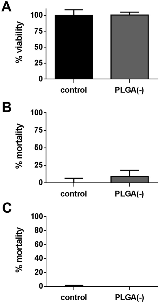
Atmospheric pollution is mainly composed of volatile pollutants and particulate matter that strongly interact. However, their specific roles in the induction of cellular toxicity, in particular the impact of the vectorization of atmospheric pollutants by ultrafine particles, remains to be fully elucidated. For this purpose, non-toxic poly-lactic co-glycolic acid (PLGA) nanoparticles were synthesized and three pollutants (benzo(a)pyrene, naphthalene and di-ethyl-hexyl-phthalate) were adsorbed on the surface of the nanoparticles in order to evaluate the toxicity (cytotoxicity, genotoxicity and ROS induction) of these complexes to a human airway epithelial cell line. The adsorption of the pollutants onto the nanoparticles was confirmed by HPLC analysis. Interestingly, the cytotoxicity assays (MTT, LDH and CellTox Green) clearly demonstrated that the vectorization by nanoparticles decreases the toxicity of the adsorbed pollutants. Genotoxicity was assessed by the micronucleus test and the comet assay and showed no increase in primary DNA damage or in chromosomal aberrations of nanoparticle vectorized pollutants. Neither cytotoxicity nor genotoxicity was correlated with ROS induction. To conclude, our results indicate that the vectorization of pollutants by nanoparticles does not potentiate the toxicity of the pollutants studied and that, on the contrary, adsorption onto nanoparticles could protect cells against pollutants' toxicity.


Conflict of interest statement

Competing Interests: The authors have declared that no competing interests exist.

Figures

Fig 1
Fig 1. Evaluation of the cytotoxicity of PLGA(-) nanoparticles.
H292 cells were treated for 15h with 250μg.ml-1 of PLGA(-) nanoparticles and cytotoxicity was assessed by MTT assay (A), LDH assay (B) and CellTox green assay® (C) and are respectively presented as percentage of viability (A) and mortality (B and C).
Fig 2
Fig 2. Cytotoxicity of free versus PLGA-adsorbed naphthalene.
H292 cells were treated with 64.1μg.ml-1 (500μM) of free (NA) or nanoparticle-adsorbed naphthalene (PLGA(-) NA) before assessing their viability by MTT assay (A, C, E) and their mortality by LDH assay (B, D) or CellTox Green® assay (F), following three protocols: a 3-hour treatment associated with S9 metabolic activation followed by a 15-hour recovery period (S9+ 3h/15h: A-B), a 3-hour treatment followed by a 15-hour recovery period (S9- 3h/15h: C-D) and a 15-hour treatment (E-F). T-test *: p<0.05; **: p<0.01; ***: p<0.005 versus free naphthalene.
Fig 3
Fig 3. ROS induction by free versus PLGA-adsorbed naphthalene.
H292 cells were treated for 5 hours with 64.1μg.ml-1 (500μM) of free (NA) or nanoparticle-adsorbed naphthalene (PLGA(-) NA) before measuring the ROS induction by the H2DCF-DA method. T-test *: p<0.05; ***: p<0.005 versus free naphthalene; §§: p<0.01 versus control.
Fig 4
Fig 4. Genotoxicity of free versus PLGA-adsorbed naphthalene.
H292 cells were treated with 0 to 500μM (64.1μg.ml-1) of free (-) or nanoparticle-adsorbed naphthalene (PLGA(-))before assessing the genotoxicity. DNA damage was evaluated by the comet assay (A) and the modified comet assays with the human 8-Oxo-guanine-glycosylase 1 (B). DNA damage is expressed as the percentage of DNA in the tail. Chromosomal aberration was determined by the micronucleus test (C). +: positive control. Mann—Whitney U-test (A, B) *: p<0.05 versus the negative control; Chi2 test (C) *:p<0.05 versus the negative control.
Fig 5
Fig 5. Cytotoxicity of free versus PLGA-adsorbed benzo(a)pyrene.
H292 cells were treated with 12.6μg.ml-1 (50μM) of free (B(a)P) or nanoparticle-adsorbed benzo(a)pyrene (PLGA(-) B(a)P) before assessing the viability by MTT assay (A, C, E) and the mortality by LDH assay (B, D) following three protocols: a 3-hour treatment associated with S9 metabolic activation followed by a 15-hour recovery period (S9+ 3h/15h: A-B), a 3-hour treatment followed by a 15-hour recovery period (S9- 3h/15h: C-D) and a 15-hour treatment (E). T-test *: p<0.05; **: p<0.01; ***: p<0.005 versus free benzo(a)pyrene.
Fig 6
Fig 6. ROS induction by free versus PLGA-adsorbed benzo(a)pyrene.
H292 cells were treated for 5 hours with 12.6μg.ml-1 (50μM) of free (B(a)P) or nanoparticle-adsorbed benzo(a)pyrene (PLGA(-) B(a)P) before measuring the ROS induction by the H2DCF-DA method. T-test **: p<0.01; ***: p<0.005 versus free benzo(a)pyrene.
Fig 7
Fig 7. Genotoxicity of free versus PLGA-adsorbed benzo(a)pyrene.
H292 cells were treated with 0 to 50μM (12.6μg.ml-1) of free (-) or nanoparticle-adsorbed benzo(a)pyrene (PLGA(-))before assessing the genotoxicity. DNA damage was evaluated by the comet assay (A) and the modified comet assays with the human 8-Oxo-guanine-glycosylase 1 (B). DNA damage is expressed as the percentage of DNA in the tail. Chromosomal aberration was determined by the micronucleus test (C). +: positive control. Mann—Whitney U-test (A, B) *: p<0.05 versus the negative control; Chi2 test (C) *:p<0.05 versus the negative control.
Fig 8
Fig 8. Cytotoxicity of free versus PLGA-adsorbed di-ethyl-hexyl phthalate (DEHP).
H292 cells were treated for 15 hours with 58.6μg.ml-1 (150μM) of free (B(a)P) or nanoparticle-adsorbed benzo(a)pyrene (PLGA(-) B(a)P) before assessing the viability by MTT assay (A) and the mortality by CellTox Green® assay (B). T-test ***: p<0.005 versus free DEHP.
Fig 9
Fig 9. ROS induction by free versus PLGA-adsorbed di-ethyl-hexyl phthalate (DEHP).
H292 cells were treated for 5 hours with 58.6μg.ml-1 (150μM) of free (DEHP) or nanoparticle-adsorbed pollutant (PLGA(-) DEHP) before measuring the ROS induction by the H2DCF-DA method. T-test no significant versus free DEHP.
Fig 10
Fig 10. Genotoxicity of free versus PLGA-adsorbed di-ethyl-hexyl phthalate (DEHP).
H292 cells were treated with 0 to 150μM (58.6μg.ml-1) of free (-) or nanoparticle-adsorbed pollutant (PLGA(-) DEHP) before assessing the genotoxicity. DNA damage was evaluated by the comet assay (A) and the modified comet assays with the human 8-Oxo-guanine-glycosylase 1 (B). DNA damage is expressed as the percentage of DNA in the tail. Chromosomal aberration was determined by the micronucleus test (C). +: positive control. Mann—Whitney U-test (A, B) *: p<0.05 versus the negative control; Chi2 test (C) *:p<0.05 versus the negative control.

References

    1. Taylor DA. Dust in the wind. Environmental health perspectives. 2002;110(2):A80–7. . - PMC - PubMed
    1. Ris C. U.S. EPA health assessment for diesel engine exhaust: a review. Inhalation toxicology. 2007;19 Suppl 1:229–39. doi: 10.1080/08958370701497960 - DOI - PubMed
    1. Westerdahl D, Fruin S, Sax T, Fine PM, Sioutas C. Mobile platform measurements of ultrafine particles and associated pollutant concentrations on freeways and residential streets in Los Angeles. Atmospheric Environment. 2005;39(20):3597–610. http://dx.doi.org/10.1016/j.atmosenv.2005.02.034. - DOI
    1. Sioutas C, Delfino RJ, Singh M. Exposure assessment for atmospheric ultrafine particles (UFPs) and implications in epidemiologic research. Environmental health perspectives. 2005;113(8):947–55. doi: 10.1289/ehp.7939 . - DOI - PMC - PubMed
    1. Valavanidis A, Fiotakis K, Vlachogianni T. Airborne particulate matter and human health: toxicological assessment and importance of size and composition of particles for oxidative damage and carcinogenic mechanisms. Journal of environmental science and health Part C, Environmental carcinogenesis & ecotoxicology reviews. 2008;26(4):339–62. doi: 10.1080/10590500802494538 - DOI - PubMed

LinkOut - more resources