. 2017 Aug 1;58(10):4390–4398.
doi: 10.1167/iovs.17-21861.

Dynamic Imaging of the Eye, Optic Nerve, and Extraocular Muscles With Golden Angle Radial MRI

Affiliations

Dynamic Imaging of the Eye, Optic Nerve, and Extraocular Muscles With Golden Angle Radial MRI

Saikat Sengupta et al. Invest Ophthalmol Vis Sci. .

Erratum in

  • Erratum.
    [No authors listed] [No authors listed] Invest Ophthalmol Vis Sci. 2017 Sep 1;58(11):4799. doi: 10.1167/iovs.17-22948a. Invest Ophthalmol Vis Sci. 2017. PMID: 28973336 Free PMC article. No abstract available.

Abstract

Purpose: The eye and its accessory structures, the optic nerve and the extraocular muscles, form a complex dynamic system. In vivo magnetic resonance imaging (MRI) of this system in motion can have substantial benefits in understanding oculomotor functioning in health and disease, but has been restricted to date to imaging of static gazes only. The purpose of this work was to develop a technique to image the eye and its accessory visual structures in motion.

Methods: Dynamic imaging of the eye was developed on a 3-Tesla MRI scanner, based on a golden angle radial sequence that allows freely selectable frame-rate and temporal-span image reconstructions from the same acquired data set. Retrospective image reconstructions at a chosen frame rate of 57 ms per image yielded high-quality in vivo movies of various eye motion tasks performed in the scanner. Motion analysis was performed for a left-right version task where motion paths, lengths, and strains/globe angle of the medial and lateral extraocular muscles and the optic nerves were estimated.

Results: Offline image reconstructions resulted in dynamic images of bilateral visual structures of healthy adults in only ∼15-s imaging time. Qualitative and quantitative analyses of the motion enabled estimation of trajectories, lengths, and strains on the optic nerves and extraocular muscles at very high frame rates of ∼18 frames/s.

Conclusions: This work presents an MRI technique that enables high-frame-rate dynamic imaging of the eyes and orbital structures. The presented sequence has the potential to be used in furthering the understanding of oculomotor mechanics in vivo, both in health and disease.


Figures
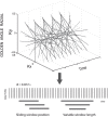
Figure 1
Figure 1
The golden angle radial imaging sequence and image reconstruction. Data profiles spaced by 111.246° are acquired continuously in time. The profiles can be grouped retrospectively to produce an image at arbitrary time positions and with arbitrary time window widths. Image can be reconstructed in a sliding window format with fast frame rates for videos of moving anatomic structures.
Figure 2
Figure 2
Axial and sagittal T2-weighted scout images used for planning the imaging slices. Manually landmarked points on the dynamic images for motion analysis are shown here on the axial T2w image. Lateral EOMs: blue points, optic nerves: red points, medial EOMs: green points, annulus of Zinn: white points, lens: orange points. EOM lengths were estimated from second-order polynomial fits to blue (lateral) and green (medial) points. Optic nerve lengths were estimated from fits to red points. The globe angle was estimated as the acute angle between the line segments joining the lens and the optic nerve head, and the optic nerve head and the AOZ (black and white lines in the right eye).
Figure 3
Figure 3
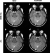
Images from subject 1 at rest and fixation at time points 360 ms and 2 seconds. Eye and optic nerve drift is observed in the rest images.
Figure 4
Figure 4
(a, b) Time frame images 0.5 s apart of left–right version task in two subjects. (c, d) Second-order polynomial fits to the landmarked point of insertion of the globes. Blue: lateral EOMs, red: optic nerves, green: medial EOMs. Motion tracks are displayed on top of the last frame of the dextroversion (c) and levoversion (d) task.
Figure 5
Figure 5
Dynamic length and angle data over the 2.85 seconds of a left–right version cycle. Circles represent lengths and angles estimated from manually selected points. Solid lines are polynomial fits to the points. Ten scans from subjects are shown. (a, e) Right and left lateral EOM lengths. Black and gray arrowheads show data from the two scans of subject 1 and subject 2, respectively. The lengths estimated from the repeated scans match closely. (b, f) Right and left medial EOM lengths. (c, g) Right and left optic nerve lengths. (d, h) Right and left globe angles.
Figure 6
Figure 6
Time frame sagittal images 0.5 s apart of up–down version tasks in two subjects.
Figure 7
Figure 7
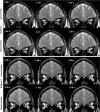
Coronal plane images of levoversion (top) and dextroversion (bottom) in two subjects. Action of all four EOMs, optic nerves, and superior oblique muscles can be observed in the motion (also see corresponding Supplementary Videos).
Figure 8
Figure 8
Images reconstructed from 4, 16, 32, 64, 96, 128, 192, and 256 profiles per image. Larger number of profiles improves image detail and SNR, at the cost of a larger temporal window, which can blur fast-moving structures. This flexibility of retrospective image reconstruction is provided only by the golden angle sequence.

References

    1. Eggert T. . Eye movement recordings: methods. Dev Ophthalmol. 2007; 40: 15– 34. - PubMed
    1. Miller JM, Robinson DA. . A model of the mechanics of binocular alignment. Comput Biomed Res. 1984; 17: 436– 470. - PubMed
    1. Demer JL. . Mechanics of the orbita. Dev Ophthalmol. 2007; 40: 132– 157. - PMC - PubMed
    1. Kono R, Clark RA, Demer JL. . Active pulleys: magnetic resonance imaging of rectus muscle paths in tertiary gazes. Invest Ophthalmol Vis Sci. 2002; 43: 2179– 2188. - PubMed
    1. Schutte S, van den Bedem SP, van Keulen F, van der Helm FC, Simonsz HJ. . A finite-element analysis model of orbital biomechanics. Vision Res. 2006; 46: 1724– 1731. - PubMed

Publication types