

Targeting Phospholipase D4 Attenuates Kidney Fibrosis
- PMID: 28814511
- PMCID: PMC5698063
- DOI: 10.1681/ASN.2016111222
Targeting Phospholipase D4 Attenuates Kidney Fibrosis
Abstract
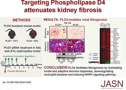
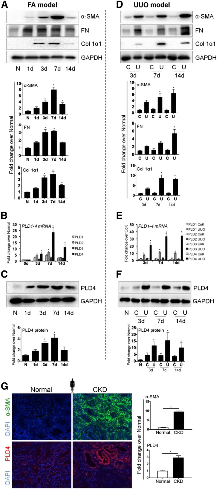
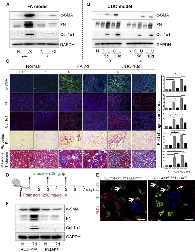
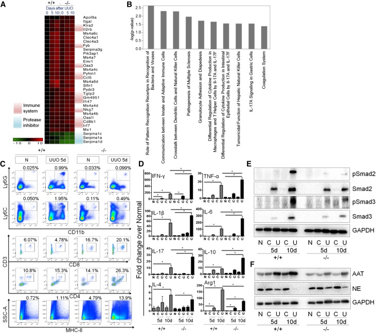
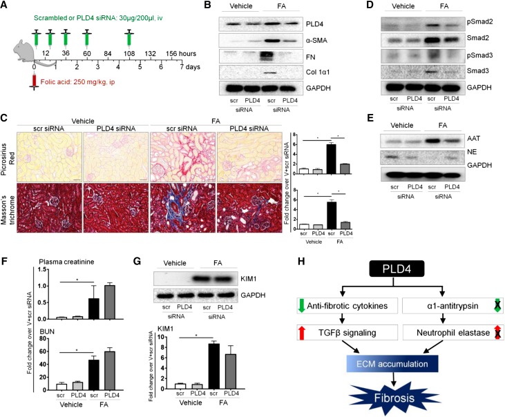
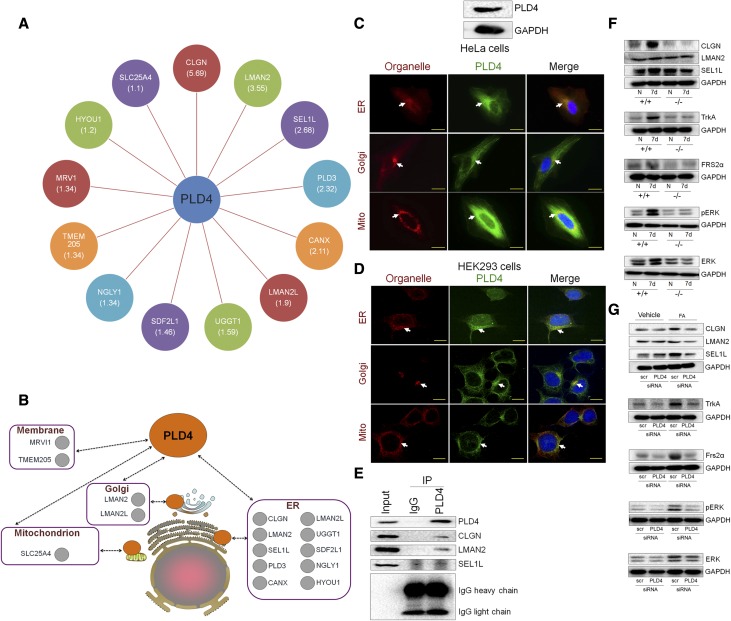
Phospholipase D4 (PLD4), a single-pass transmembrane glycoprotein, is among the most highly upregulated genes in murine kidneys subjected to chronic progressive fibrosis, but the function of PLD4 in this process is unknown. Here, we found PLD4 to be overexpressed in the proximal and distal tubular epithelial cells of murine and human kidneys after fibrosis. Genetic silencing of PLD4, either globally or conditionally in proximal tubular epithelial cells, protected mice from the development of fibrosis. Mechanistically, global knockout of PLD4 modulated innate and adaptive immune responses and attenuated the upregulation of the TGF-β signaling pathway and α1-antitrypsin protein (a serine protease inhibitor) expression and downregulation of neutrophil elastase (NE) expression induced by obstructive injury. In vitro, treatment with NE attenuated TGF-β-induced accumulation of fibrotic markers. Furthermore, therapeutic targeting of PLD4 using specific siRNA protected mice from folic acid-induced kidney fibrosis and inhibited the increase in TGF-β signaling, decrease in NE expression, and upregulation of mitogen-activated protein kinase signaling. Immunoprecipitation/mass spectrometry and coimmunoprecipitation experiments confirmed that PLD4 binds three proteins that interact with neurotrophic receptor tyrosine kinase 1, a receptor also known as TrkA that upregulates mitogen-activated protein kinase. PLD4 inhibition also prevented the folic acid-induced upregulation of this receptor in mouse kidneys. These results suggest inhibition of PLD4 as a novel therapeutic strategy to activate protease-mediated degradation of extracellular matrix and reverse fibrosis.
Keywords: TGF-beta; chronic kidney disease; fibrosis; immunology.
Copyright © 2017 by the American Society of Nephrology.
Figures
Comment in
-
Chronic kidney disease: PLD4 regulates kidney fibrosis.Nat Rev Nephrol. 2017 Oct;13(10):597. doi: 10.1038/nrneph.2017.125. Epub 2017 Sep 4. Nat Rev Nephrol. 2017. PMID: 28869252 No abstract available.
Similar articles
-
A single-domain i-body, AD-114, attenuates renal fibrosis through blockade of CXCR4.JCI Insight. 2022 Feb 22;7(4):e143018. doi: 10.1172/jci.insight.143018. JCI Insight. 2022. PMID: 35015734 Free PMC article.
-
Connective tissue growth factor induces renal fibrosis via epidermal growth factor receptor activation.J Pathol. 2018 Feb;244(2):227-241. doi: 10.1002/path.5007. Epub 2018 Jan 10. J Pathol. 2018. PMID: 29160908
-
Phospholipase D family member 4, a transmembrane glycoprotein with no phospholipase D activity, expression in spleen and early postnatal microglia.PLoS One. 2010 Nov 11;5(11):e13932. doi: 10.1371/journal.pone.0013932. PLoS One. 2010. PMID: 21085684 Free PMC article.
-
Targeting TGF-β Signaling in Kidney Fibrosis.Int J Mol Sci. 2018 Aug 27;19(9):2532. doi: 10.3390/ijms19092532. Int J Mol Sci. 2018. PMID: 30150520 Free PMC article. Review.
-
Defining therapeutic targets for renal fibrosis: Exploiting the biology of pathogenesis.Biomed Pharmacother. 2021 Nov;143:112115. doi: 10.1016/j.biopha.2021.112115. Epub 2021 Sep 3. Biomed Pharmacother. 2021. PMID: 34488081 Review.
Cited by
-
Mechanism of Chaihuang-Yishen formula to attenuate renal fibrosis in the treatment of chronic kidney disease: Insights from network pharmacology and experimental validation.Heliyon. 2024 Aug 8;10(16):e35728. doi: 10.1016/j.heliyon.2024.e35728. eCollection 2024 Aug 30. Heliyon. 2024. PMID: 39220918 Free PMC article.
-
MicroRNA miR-145-5p inhibits Phospholipase D 5 (PLD5) to downregulate cell proliferation and metastasis to mitigate prostate cancer.Bioengineered. 2021 Dec;12(1):3240-3251. doi: 10.1080/21655979.2021.1945361. Bioengineered. 2021. PMID: 34238129 Free PMC article.
-
A novel lipid metabolism gene signature for clear cell renal cell carcinoma using integrated bioinformatics analysis.Front Cell Dev Biol. 2023 Feb 14;11:1078759. doi: 10.3389/fcell.2023.1078759. eCollection 2023. Front Cell Dev Biol. 2023. PMID: 36866272 Free PMC article.
-
Discovery of PLD4 modulators by high-throughput screening and kinetic analysis.Results Chem. 2024 Jan;7:101349. doi: 10.1016/j.rechem.2024.101349. Epub 2024 Feb 8. Results Chem. 2024. PMID: 38560090 Free PMC article.
-
Improved predictability of pancreatic ductal adenocarcinoma diagnosis using a blood immune cell biomarker panel developed from bulk mRNA sequencing and single-cell RNA-sequencing.Cancer Immunol Immunother. 2023 Aug;72(8):2757-2768. doi: 10.1007/s00262-023-03458-8. Epub 2023 May 10. Cancer Immunol Immunother. 2023. PMID: 37165046 Free PMC article.
References
-
- Kurts C, Panzer U, Anders HJ, Rees AJ: The immune system and kidney disease: Basic concepts and clinical implications. Nat Rev Immunol 13: 738–753, 2013 - PubMed
-
- Craciun FL, Bijol V, Ajay AK, Rao P, Kumar RK, Hutchinson J, Hofmann O, Joshi N, Luyendyk JP, Kusebauch U, Moss CL, Srivastava A, Himmelfarb J, Waikar SS, Moritz RL, Vaidya VS: RNA sequencing identifies novel translational biomarkers of kidney fibrosis. J Am Soc Nephrol 27: 1702–1713, 2016 - PMC - PubMed
-
- Yoshikawa F, Banno Y, Otani Y, Yamaguchi Y, Nagakura-Takagi Y, Morita N, Sato Y, Saruta C, Nishibe H, Sadakata T, Shinoda Y, Hayashi K, Mishima Y, Baba H, Furuichi T: Phospholipase D family member 4, a transmembrane glycoprotein with no phospholipase D activity, expression in spleen and early postnatal microglia. PLoS One 5: e13932, 2010 - PMC - PubMed
MeSH terms
Substances
Grants and funding
LinkOut - more resources
Full Text Sources
Other Literature Sources
Molecular Biology Databases
