. 2017 Aug 16;9(403):eaan2415.
doi: 10.1126/scitranslmed.aan2415.

Direct detection of early-stage cancers using circulating tumor DNA

Affiliations

Direct detection of early-stage cancers using circulating tumor DNA

Jillian Phallen et al. Sci Transl Med. .

Abstract

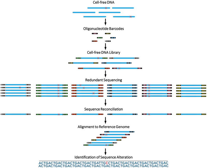
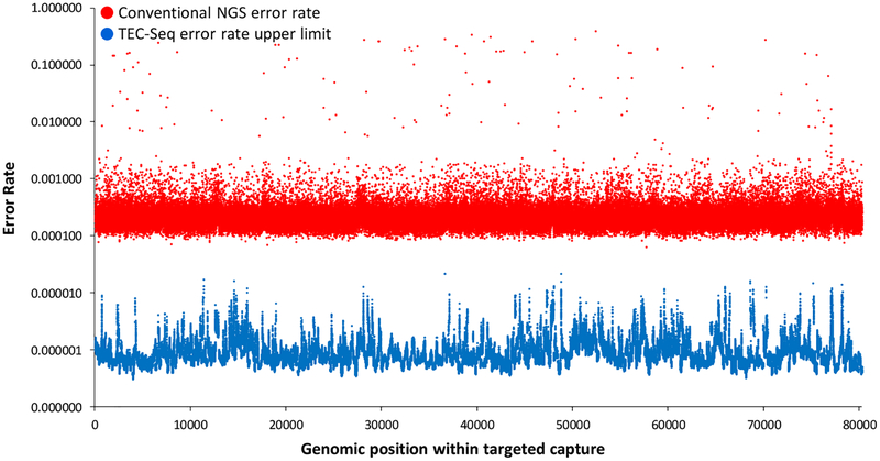
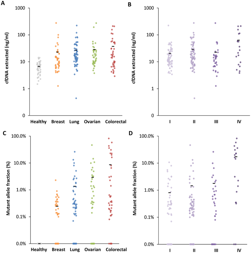
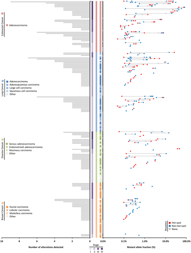
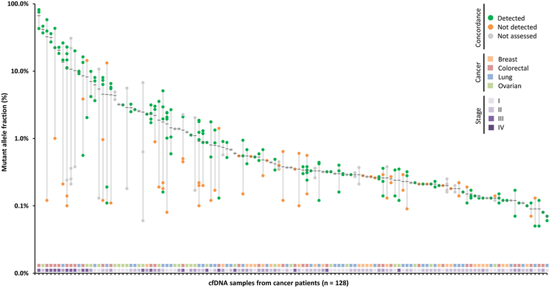
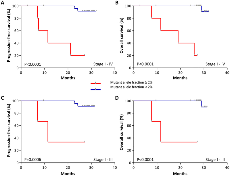
Early detection and intervention are likely to be the most effective means for reducing morbidity and mortality of human cancer. However, development of methods for noninvasive detection of early-stage tumors has remained a challenge. We have developed an approach called targeted error correction sequencing (TEC-Seq) that allows ultrasensitive direct evaluation of sequence changes in circulating cell-free DNA using massively parallel sequencing. We have used this approach to examine 58 cancer-related genes encompassing 81 kb. Analysis of plasma from 44 healthy individuals identified genomic changes related to clonal hematopoiesis in 16% of asymptomatic individuals but no alterations in driver genes related to solid cancers. Evaluation of 200 patients with colorectal, breast, lung, or ovarian cancer detected somatic mutations in the plasma of 71, 59, 59, and 68%, respectively, of patients with stage I or II disease. Analyses of mutations in the circulation revealed high concordance with alterations in the tumors of these patients. In patients with resectable colorectal cancers, higher amounts of preoperative circulating tumor DNA were associated with disease recurrence and decreased overall survival. These analyses provide a broadly applicable approach for noninvasive detection of early-stage tumors that may be useful for screening and management of patients with cancer.


Figures

Figure 1.
Figure 1.. Schematic of TEC-Seq method.
Cell-free DNA is extracted from blood and converted to a genomic library through ligation of a pool containing a small number of dual index barcode adapters. The resulting cfDNA library is captured and redundantly sequenced to produce multiple duplicates of each DNA fragment. Sequence reconciliation among duplicate fragments identifies alterations present in identical DNA molecules with the same start and end position and exogenous barcodes. Alignment to the reference genome of multiple distinct molecules containing identical redundant changes is used to identify bona fide alterations.
Figure 2.
Figure 2.. TEC-Seq error correction.
Sequencing error rates of conventional next generation sequencing and theoretical upper limit for TEC-Seq are indicated at each base in the captured regions of interest (P < 0.0001, paired t test). Error rates are determined by identifying the number of alterations at each base (or assuming one alteration per base if no error was identified) divided by the total coverage at each base among the 44 healthy individuals analyzed.
Figure 3.
Figure 3.. cfDNA and ctDNA in healthy individuals and patients with cancer.
Amount of cfDNA extracted from all heathy individuals and patients with different cancer types (ng/ml) (A) and from cancer patients of different stages (B). Mutant allele fraction (%) of ctDNA detected in heathy individuals and patients with different cancer types (C) and in cancer patients of different stages (D). Means for each group are represented by the black bars in the columns analyzed. In patients for whom multiple alterations were detected, the highest value is indicated. Clinical characteristics of patients and stages are indicated in table S3.
Figure 4.
Figure 4.. ctDNA in patients with breast, colorectal, lung, and ovarian cancer.
Patients (n = 194) are each represented by a tick mark. Left: Bar chart shows the number of alterations detected for each case. Center: Stage, cancer type, and histopathological subtype are represented by colored vertical bars. Right: Mutant allele fractions for each alteration detected per patient are indicated with an ‘x’ at the mean. Alterations are colored based on hot-spot status and whether any alterations were detected in the case.
Figure 5.
Figure 5.. Concordance between alterations in plasma and tissue.
Mutant allele fractions observed in the plasma are indicated for each alteration identified with a black bar at the mean. Presence of alterations in matched tumor specimens is indicated with green dots, whereas non-concordant alterations are indicated in orange and those not assessed in gray. Stage and cancer type for each patient are plotted in the two horizontal tracks at the bottom of the figure.
Figure 6.
Figure 6.. Pre-operative ctDNA amounts and outcome in colorectal cancer patients.
Kaplan-Meier curves depict progression-free survival (A, Log-rank test p < 0.0001) and overall survival (B, Log-rank test p < 0.0001) of 31 colorectal cancer patients, stage I - IV, stratified based on a ctDNA mutant allele fraction threshold of 2%. Kaplan-Meier analyses of the 27 patients with stage I - III disease for progression-free survival (C, Log-rank test p = 0.0006) and overall survival (D, Log-rank test p < 0.0001) were performed using the same threshold to examine the association of ctDNA with outcome in patients without stage IV disease.

References

    1. Torre LA et al., Global cancer statistics, 2012. CA Cancer J Clin 65, 87 (2015). - PubMed
    1. W. H. Organization, Guide to Cancer Early Diagnosis. Guide to Cancer Early Diagnosis, (2017).
    1. Mazzucchelli R, Colanzi P, Pomante R, Muzzonigro G, Montironi R, Prostate tissue and serum markers. Advances in clinical pathology : the official journal of Adriatic Society of Pathology 4, 111 (2000). - PubMed
    1. Ruibal Morell A, CEA serum levels in non-neoplastic disease. The International journal of biological markers 7, 160 (1992). - PubMed
    1. Galli C, Basso D, Plebani M, CA 19–9: handle with care. Clinical chemistry and laboratory medicine 51, 1369 (2013). - PubMed

Substances