. 2017 Aug 17;2(16):e90103.
doi: 10.1172/jci.insight.90103.

Rapamycin reversal of VEGF-C-driven lymphatic anomalies in the respiratory tract

Affiliations

Rapamycin reversal of VEGF-C-driven lymphatic anomalies in the respiratory tract

Peter Baluk et al. JCI Insight. .

Abstract

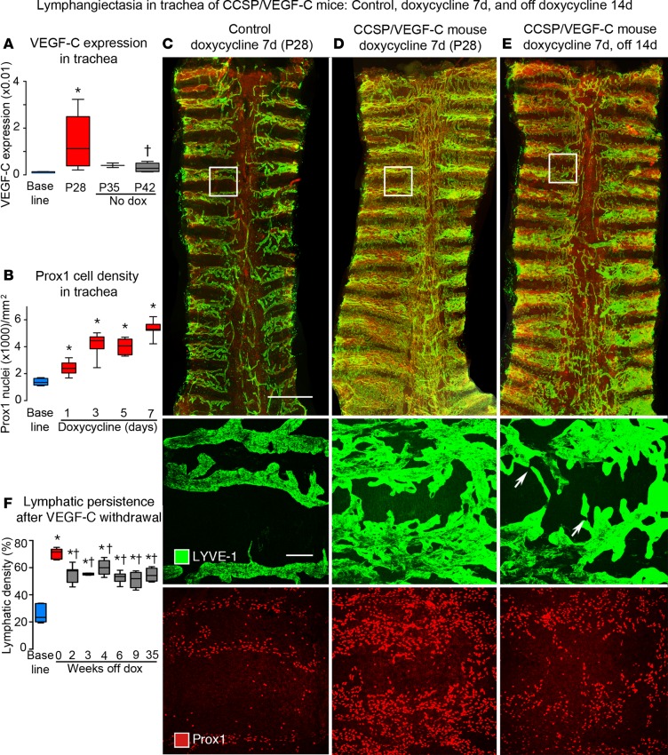
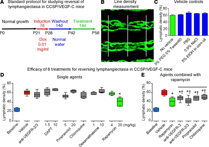
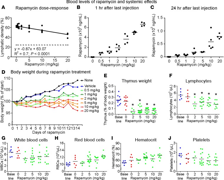
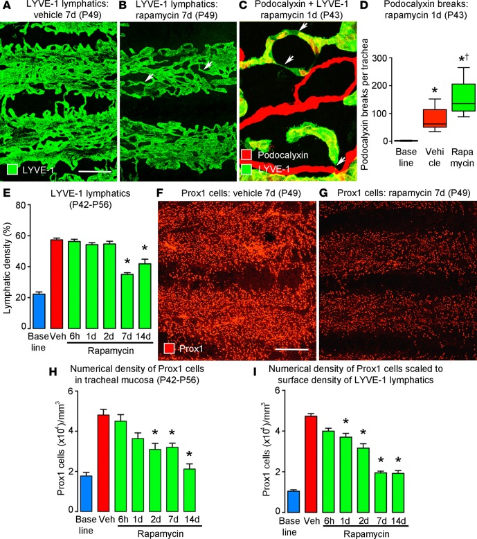
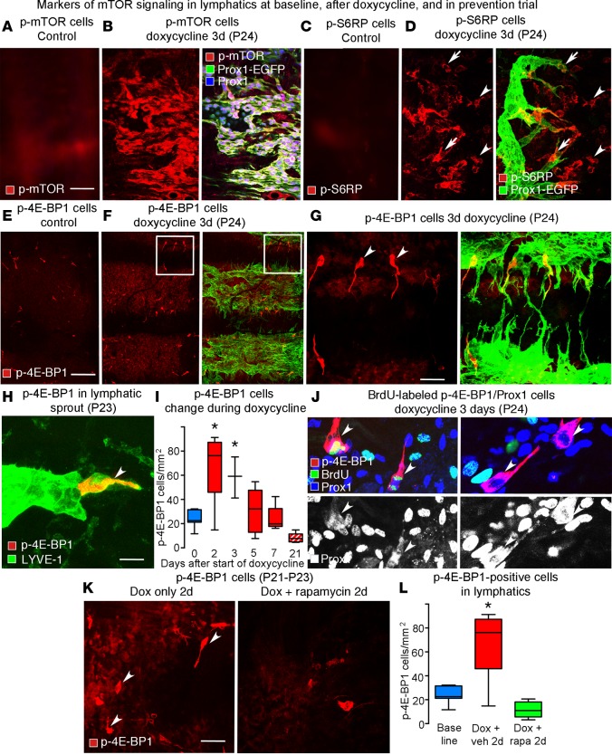
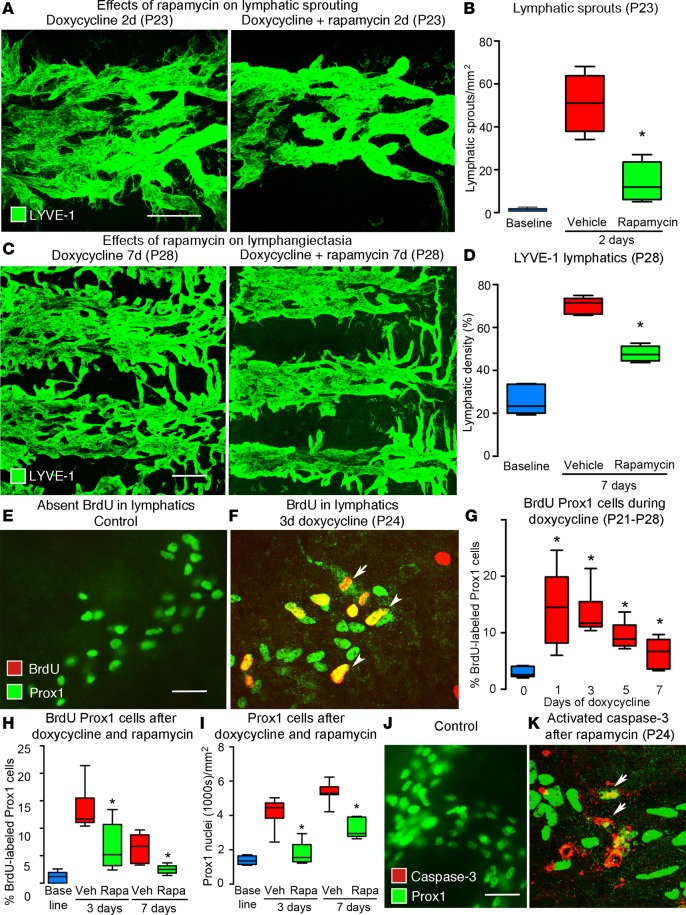
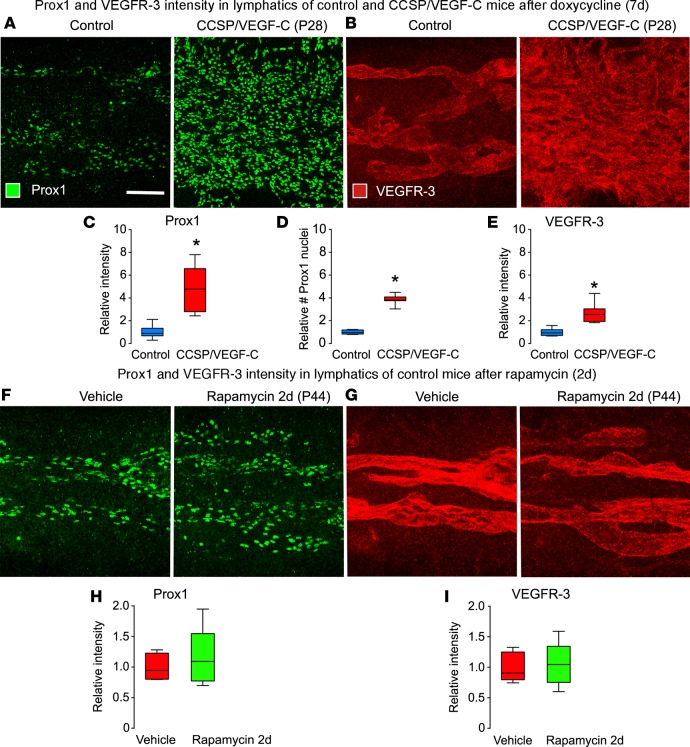
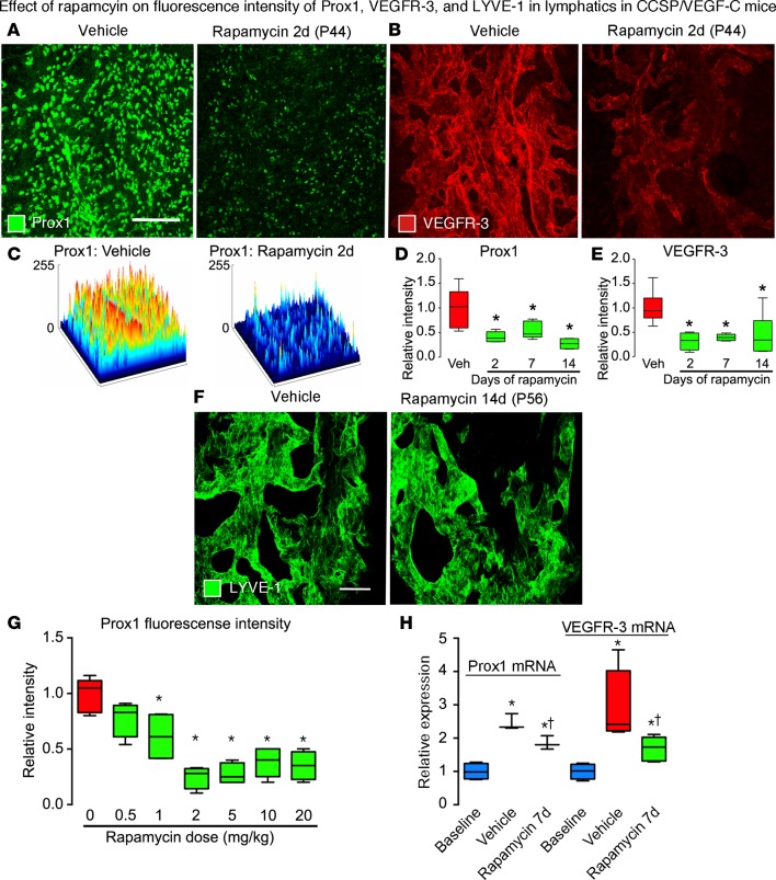
Lymphatic malformations are serious but poorly understood conditions that present therapeutic challenges. The goal of this study was to compare strategies for inducing regression of abnormal lymphatics and explore underlying mechanisms. CCSP-rtTA/tetO-VEGF-C mice, in which doxycycline regulates VEGF-C expression in the airway epithelium, were used as a model of pulmonary lymphangiectasia. After doxycycline was stopped, VEGF-C expression returned to normal, but lymphangiectasia persisted for at least 9 months. Inhibition of VEGFR-2/VEGFR-3 signaling, Notch, β-adrenergic receptors, or autophagy and antiinflammatory steroids had no noticeable effect on the amount or severity of lymphangiectasia. However, rapamycin inhibition of mTOR reduced lymphangiectasia by 76% within 7 days without affecting normal lymphatics. Efficacy of rapamycin was not increased by coadministration with the other agents. In prevention trials, rapamycin suppressed VEGF-C-driven mTOR phosphorylation and lymphatic endothelial cell sprouting and proliferation. However, in reversal trials, no lymphatic endothelial cell proliferation was present to block in established lymphangiectasia, and rapamycin did not increase caspase-dependent apoptosis. However, rapamycin potently suppressed Prox1 and VEGFR-3. These experiments revealed that lymphangiectasia is remarkably resistant to regression but is responsive to rapamycin, which rapidly reduces and normalizes the abnormal lymphatics without affecting normal lymphatics.

Keywords: Vascular Biology.


Conflict of interest statement

Conflict of interest: The authors have declared that no conflict of interest exists.

Figures

Figure 1
Figure 1. Lymphatics in tracheas of CCSP/VEGF-C mice before and after doxycycline.
(A) VEGF-C mRNA expression (relative to β-actin) in trachea increased on doxycycline and returned almost to baseline after 14-day doxycycline washout period. (B) Number of Prox1-positive nuclei before and during doxycycline administration. (C–E) Overview of tracheal whole mounts stained for LYVE-1 (green) and Prox1 (red) from mice given 0.01 mg/ml doxycycline for 7 days (P21–P28) and perfused at P28 (C and D) or after 14-day washout period (P42) (E). (C) Trachea from a control (single-transgenic) mouse, with normal lymphatics restricted to intercartilage spaces and over the trachealis muscle at the center. (D) Trachea from a double-transgenic CCSP/VEGF-C mouse after doxycycline for 7 days, where lymphatics cover a much larger area and increase in abundance toward the caudal end. (E) Trachea from a P42 double-transgenic mouse 14 days after doxycycline withdrawal; lymphatics are less numerous than at P28, especially in central muscular region, but most of the lymphatic abnormality persists. Boxed regions are shown at higher magnification below. The middle row shows the boxed region stained for LYVE-1 showing normal lymphatics (C), lymphangiectasia at P28 (D), and spontaneous regression between P28 and P42 (E). Some lymphatics are narrowed regions (arrows). The lower row shows Prox1-positive nuclei at the beginning (C) and end of doxycycline treatment (D) and 14 days after withdrawal (E). Scale bar: 1 millimeter (top row); 50 μm (middle and bottom rows). (F) Extent of LYVE-1 lymphatics in trachea before doxycycline (blue box), after doxycycline (red box), and after doxycycline withdrawal (gray boxes). Values show the lymphatic expansion during doxycycline and some regression during the 2-week withdrawal period (P28–P42) but none thereafter throughout the 9-month study period (gray boxes were not significantly different from each other). n = 3 to 10 mice/group. *P < 0.05 vs. baseline group, P < 0.05 vs. P28, ANOVA. Box and whisker plots show the median, first and third quartiles, and maximum and minimum.
Figure 2
Figure 2. Standard protocol and comparison of effects of six treatment strategies.
(A) Standard protocol for studying effects of treatment on lymphangiectasia in CCSP/VEGF-C mice on doxycycline (0.01 mg/ml) for 7 days, off doxycycline for 14 days, and then treated for 14 days (n = 4–6 mice/group). (B) Approach for measuring line density of tracheal lymphatics stained for LYVE-1 by overlaying 3 parallel lines and assessing the fraction of pixels with a brightness of 100 or greater (on a scale of 0–255) with ImageJ. (C) Density of tracheal lymphatics in CCSP/VEGF-C mice (P42–P56) after treatment with one of several vehicles used for the active therapeutics. (D) Lymphatic density in tracheas of CCSP/VEGF-C mice treated in the standard protocol with doxycycline (P21–P28), off doxycycline (P28–P42), and treated with one of six therapeutics for 14 days (P42–P56). Only the high dose of rapamycin (green box, 20 mg/kg) induced significant regression compared with the vehicle-treated group (red box). Treatment with lower doses of candidate therapeutics had no significant effect. (E) Effects of treatment with rapamycin in combination with other agents (P42–P56). The amount of lymphatic regression was not significantly greater (†) than that found after rapamycin alone (green box) but was less than that after treatment with vehicle (*P < 0.05 vs. vehicle, ANOVA). Box and whisker plots show the median, first and third quartiles, and maximum and minimum.
Figure 3
Figure 3. Dose response and assessment of toxicity of rapamycin.
Dose-response effect of rapamycin (0.5–20 mg/kg i.p. daily, P42–P56) in CCSP/VEGF-C mice for 14 days on reversal of lymphangiectasia (A). Dashed line in A marks the value for tracheal lymphatics in normal mice. Rapamycin blood level at 1 hour (B) and 24 hours (C) after last dose. Body weight (D), thymus weight (E), and blood counts for lymphocytes (F), white blood cells (G), red blood cells (H), hematocrit (I), and platelets (J) at 24 hours after final dose. The baseline value represents that for wild-type mice. *P < 0.05 vs. vehicle, ANOVA.
Figure 4
Figure 4. Time course and magnitude of rapamycin reversal of lymphangiectasia.
(A and B) Lymphatic endothelial cells, stained for LYVE-1 after treatment for 7 days (P42–P49), form a sheet-like network after treatment with vehicle (A) but are more tubular after treatment with rapamycin (B, arrows). Scale bar: 200 μm. (C) Narrowing of lymphatics (arrows) after 1-day treatment with rapamycin. (D) Number of breaks in podocalyxin staining in lymphatics per trachea, reflecting lumen closure of lymphatics after rapamycin for 1 day (P42–P43) (n = 6 mice/group). *P < 0.05 vs. baseline; P < 0.05 vs. vehicle, ANOVA. Box and whisker plots show the median, first and third quartiles, and maximum and minimum. (E) Time course of lymphatic regression induced by rapamycin, as assessed by the extent of LYVE-1 staining in confocal microscopic projections of the tracheal surface. (F and G) Lymphatic endothelial cells stained for Prox1 after treatment with vehicle or rapamycin for 7 days (P42–P49). (H) Time course of lymphatic regression induced by rapamycin, as assessed by the number of Prox1-positive nuclei in confocal projections of tracheal surface. (I) Prox1-positive nuclei per cubic millimeter of trachea, calculated as the product of E and H. *P < 0.05 vs. vehicle (red), ANOVA, n = 5 mice/group (EI).
Figure 5
Figure 5. Staining for p-mTOR, p-S6RP, and p-4E-BP1 in growing lymphatics.
(A) Absence of p-mTOR staining in lymphatics of normal trachea. (B) Uniformly strong p-mTOR staining in lymphatics. The epithelium was excluded from the confocal projection of the tracheal whole mount. (C) Absence of p-S6RP staining in lymphatics of normal trachea. (D) Strong p-S6RP staining in lymphatic sprouts (arrows) in CCSP/VEGF-C mice after doxycycline (3 days). Arrowheads mark p-S6RP staining of other cell types. (E) Absence of p-4E-BP1 staining in lymphatics of normal trachea. (F) Strong p-4E-BP1 staining in a subset of lymphatic endothelial cells in CCSP/VEGF-C mice on doxycycline (3 days). (G) The boxed region in F shown at higher magnification. Arrowheads mark p-4E-BP1–positive cells. (H) Phospho-4E-BP1 staining in a lymphatic sprout (arrowhead) in CCSP/VEGF-C mice on doxycycline (2 days, P23). (I) Transient increase in number of p-4E-BP1–stained cells during doxycycline exposure (red boxes, P21–P28) and 14 days after withdrawal of doxycycline (red stripped box, P42). n = 3–10 mice/group. *P < 0.05 vs. vehicle, ANOVA. (J) BrdU labeling of p-4E-BP1–stained cells after doxycycline (3 days). BrdU labeling (green) in lymphatic endothelial cells is more abundant than p-4E-BP1 (red). Prox1 (white in bottom panels) is not restricted to the nuclei of dividing cells (arrowheads, left panels). Some p-4E-BP1–stained cells lack BrdU labeling (arrowheads, right panels). (K) Numerous p-4E-BP1–stained cells were observed after doxycycline alone (2 days, P21-P23) but not when doxycycline was given concurrently with rapamycin. (L) An increased number of p-4E-BP1–stained cells (red box) was observed after treatment with doxycycline (2 days, P21–P23). The increase was prevented when doxycycline was given concurrently with rapamycin (20 mg/kg, green box). *P < 0.05 vs. baseline, ANOVA, n = 5 mice/group. Scale bar: 50 μm (AD, G, and K); 200 μm (E and F); 10 μm (H). Box and whisker plots show the median, first and third quartiles, and maximum and minimum.
Figure 6
Figure 6. Effect of rapamycin on lymphatic sprouting and proliferation in tracheas of CCSP/VEGF-C mice.
(A) Lymphatic sprouts after doxycycline (left) but not after doxycycline plus rapamycin (right) (2d, P21–P23). (B) Measurements of lymphatic sprouts. Baseline, single-transgenic mice (P21). *P < 0.05 vs. vehicle, ANOVA. (C) Lymphangiogenesis after doxycycline (left) but not after doxycycline plus rapamycin (right) (7d, P21–P28). (D) Measurements of lymphatic density. *P < 0.05 vs. vehicle, ANOVA. (E) No BrdU uptake by lymphatics in control mouse, compared with (F) BrdU-labeled lymphatics (arrowheads) after doxycycline (3d). Dividing lymphatic endothelial cell (arrow). (G) BrdU labeling of lymphatics after doxycycline for 1–7 days (P21–P28). *P < 0.05 vs. baseline, ANOVA. (H) Rapamycin suppression of BrdU labeling after doxycycline for 3 or 7 days (P24 or P28), expressed as a percentage of Prox1-positive cells. (I) Reduction in Prox1-positive cells after doxycycline plus rapamycin for 3 days (87% reduction, P24) or 7 days (52% reduction, P28). (J) No activated caspase-3 staining in control lymphatics, compared with (K) staining of fragmented Prox1-positive cells (arrows) after doxycycline plus rapamycin (3d, P21 to P24). n = 5 mice/group. Scale bar: 100 μm (A); 200 μm (C); 20 μm (E, F, J, and K). Box and whisker plots show the median, first and third quartiles, and maximum and minimum.
Figure 7
Figure 7. Effects of doxycycline and rapamycin on Prox1 and VEGFR-3 in lymphatics of normal mice.
(A and B) Confocal micrographs comparing Prox1 (A) and VEGFR-3 (B) staining of lymphatics in the trachea of control mouse (left, single-transgenic CCSP) and a CCSP/VEGF-C mouse (right) after doxycycline (7 days, P21–28). Images were photographed with matching brightness and contrast settings. (C–E) Intensity of Prox1 fluorescence (C), number of Prox1-positive nuclei (D), and intensity of VEGFR-3 fluorescence (E) under same conditions as in A and B. *P < 0.05 vs. single-transgenic control, ANOVA; n = 5–6 mice/group. (F–I) Confocal micrographs comparing Prox1 (F) and VEGFR-3 (G) fluorescence in lymphatics and corresponding measurements (H and I) in normal mice after treatment with vehicle or rapamycin (2 days, P21–P23). Scale bar: 100 μm. Box and whisker plots show the median, first and third quartiles, and maximum and minimum.
Figure 8
Figure 8. Effects of rapamycin on Prox1, VEGFR-3, and LYVE-1 in lymphatics of CCSP/VEGF-C mice.
(A and B) Confocal micrographs comparing Prox1 (A) and VEGFR-3 (B) immunofluorescence in tracheas of CCSP/VEGF-C mice treated with vehicle or rapamycin (2 days, P42–P44). Images photographed with matching brightness and contrast settings. (C) Surface contour plots of intensity of Prox1 fluorescence in A after treatment with vehicle or rapamycin. Prox1 (D) and VEGFR-3 (E) fluorescence intensities in CCSP/VEGF-C mice treated, as in A and B. (F) Confocal micrographs of LYVE-1 immunofluorescence in tracheas of CCSP/VEGF-C mice, showing no difference in intensity after treatment with vehicle or rapamycin (14 days, P42–P56). (G) Rapamycin dose-response effect on Prox1 fluorescence intensity. *P < 0.05 vs. vehicle, ANOVA, n = 4–6 mice/group. (H) qRT-PCR measurements of Prox1 and VEGFR-3 expression in tracheas of CCSP/VEGF-C mice on vehicle or rapamycin (7 days, P42–P49). *P < 0.05 vs. baseline, P < 0.05 vs. vehicle, ANOVA, n = 3–6 mice/group. Scale bars: 200 μm (A and B); 50 μm (F). Box and whisker plots show the median, first and third quartiles, and maximum and minimum.

References

    1. Wassef M, et al. Vascular anomalies classification: recommendations from the International Society for the Study of Vascular Anomalies. Pediatrics. 2015;136(1):e203–e214. doi: 10.1542/peds.2014-3673. - DOI - PubMed
    1. Brouillard P, Boon L, Vikkula M. Genetics of lymphatic anomalies. J Clin Invest. 2014;124(3):898–904. doi: 10.1172/JCI71614. - DOI - PMC - PubMed
    1. Trenor CC, Chaudry G. Complex lymphatic anomalies. Semin Pediatr Surg. 2014;23(4):186–190. doi: 10.1053/j.sempedsurg.2014.07.006. - DOI - PubMed
    1. Adams DM, et al. Efficacy and safety of sirolimus in the treatment of complicated vascular anomalies. Pediatrics. 2016;137(2):e20153257. doi: 10.1542/peds.2015-3257. - DOI - PMC - PubMed
    1. Virchow R. Gesammelte Abhandlungen zur wissenschaftlichen Medicin. Frankfurt-am-Main, Germany: Verlag Meidinger Sohn & Comp.; 1856;983. https://archive.org/details/b21462161 Accessed July 18, 2017.