. 2017 Aug 16;8(1):268.
doi: 10.1038/s41467-017-00296-y.

Spatial genomic heterogeneity in multiple myeloma revealed by multi-region sequencing

Affiliations

Spatial genomic heterogeneity in multiple myeloma revealed by multi-region sequencing

L Rasche et al. Nat Commun. .

Abstract

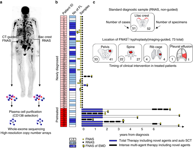
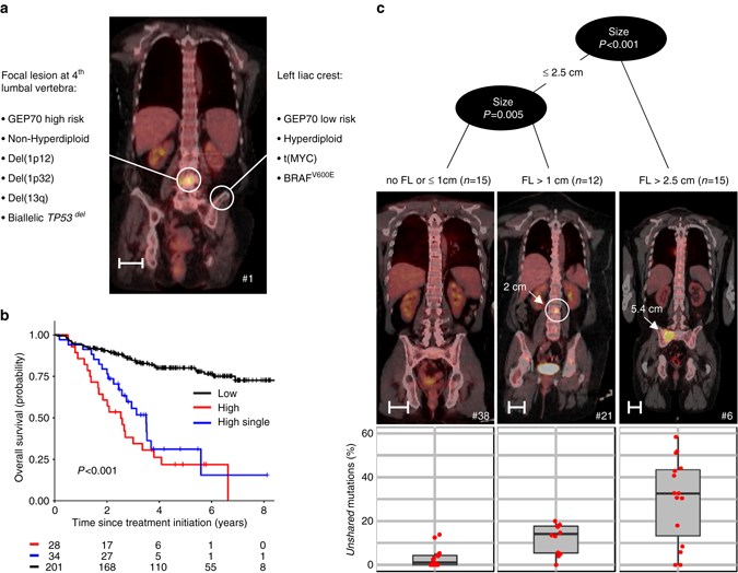
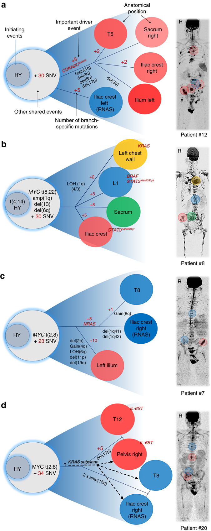
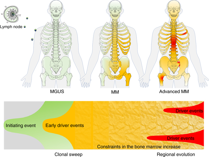
In multiple myeloma malignant plasma cells expand within the bone marrow. Since this site is well-perfused, a rapid dissemination of "fitter" clones may be anticipated. However, an imbalanced distribution of multiple myeloma is frequently observed in medical imaging. Here, we perform multi-region sequencing, including iliac crest and radiology-guided focal lesion specimens from 51 patients to gain insight into the spatial clonal architecture. We demonstrate spatial genomic heterogeneity in more than 75% of patients, including inactivation of CDKN2C and TP53, and mutations affecting mitogen-activated protein kinase genes. We show that the extent of spatial heterogeneity is positively associated with the size of biopsied focal lesions consistent with regional outgrowth of advanced clones. The results support a model for multiple myeloma progression with clonal sweeps in the early phase and regional evolution in advanced disease. We suggest that multi-region investigations are critical to understanding intra-patient heterogeneity and the evolutionary processes in multiple myeloma.In multiple myeloma, malignant cells expand within bone marrow. Here, the authors use multi-region sequencing in patient samples to analyse spatial clonal architecture and heterogeneity, providing novel insight into multiple myeloma progression and evolution.


Conflict of interest statement

B.B. is a co-inventor on patents and patent applications related to use of GEP in cancer medicine that have been licensed to Signal Genetics Inc. The remaining authors declare no conflict of interests.

Figures

Fig. 1
Fig. 1
Sample origin and processing. a Traditional iliac crest specimens (random aspirates) and CT-guided fine needle aspirates were enriched for malignant plasma cells using CD138-positive selection. Mutational and chromosomal profiles were investigated using whole-exome sequencing and high-resolution copy number arrays. b Disease state at sampling, treatment history, the total number of focal lesions in medical imaging, and the number and type of investigated samples are depicted. c Origin and number of samples. FL focal lesion, FNAS fine needle aspirate, RNAS random aspirate, EMD extramedullary disease, auto SCT autologous stem cell transplantation
Fig. 2
Fig. 2
Spatial heterogeneity in baseline and treated patients. In the upper panel of a the number of unshared copy number aberrations between paired iliac crest and fine needle aspirates (Δ CNA, > 1 Mb) is shown. In the lower panel of a heterogeneity involving the initiating events hyperdiploidy and recurrent IgH translocations as well as commonly used prognostic markers is presented with light and dark red indicating shared and unshared events, respectively. The total number of non-silent mutations is presented in b. c Proportion of clonal unshared (cancer clonal fraction (CCF) ≥ 0.8; dark blue), subclonal unshared (CCF < 0.8; cornflower blue) and shared-diff (green) non-silent mutations. In d, non-silent mutations and deletions affecting the most frequently mutated genes in our set and other recurrently mutated genes in multiple myeloma are shown. Dark blue and cornflower blue denote clonal and subclonal unshared mutations, green shared-diff mutations, red deletions and black bi-allelic events. Light colors or crossed boxes indicate shared events
Fig. 3
Fig. 3
Spatial heterogeneity: example, impact on outcome and association with the size of focal lesions. In a, unshared key drivers and the risk status in paired samples of patient no. 1 are shown. b Overall survival after enrollment into Total Therapy for 263 patients stratified by the GEP70 risk status in a routinely collected iliac crest sample and a paired CT-guided fine needle aspirate. Whereas black and red indicate patients with GEP70 low- and high-risk in both samples, respectively, the blue line shows the outcome for patients with discrepancies for paired samples (single-site high-risk). In c, recursive partitioning with the maximum size of focal lesions as the predictor of the proportion of unshared mutations was performed. Representative PET-CT images for the three size-groups are depicted (length of scale bars = 5 cm)
Fig. 4
Fig. 4
Multi-regional evolution. Cases with availability of multiple CT-guided samples were selected to analyze the phylogenetic relationship of clones from different regions. The location of samples is marked in the medical images in the right panel using the color code that was assigned to clones (left panel). The letter R indicates the right side of the body. a Four focal lesions (FL) showed similar genomic profiles, sharing the high-risk events bi-allelic deletion of CDKN2C, gain(1q) and del(17p). In contrast, a randomly biopsied non-FL site was low-risk and showed none of these events. b FLs displayed different genomic profiles with each of them containing unique driver mutations (BRAF, KRAS, and STAT3). Clones at the iliac crest and L1 contained different STAT3 mutations (STAT3Asn553Lys vs. STAT3Asp663Tyr). c Clones at T8 and the left iliac crest shared a missense NRAS mutation. Compared to the NRAS branch, the right iliac crest FL showed multiple site-specific CNAs and non-silent mutations. d Two major clones occupied four distant sites with the first clone being characterized by a non-ubiquitous IL6ST mutation. Moreover, an independent minor subclone with a missense KRAS mutation infiltrated both sites in the pelvis and T8, but not T12
Fig. 5
Fig. 5
Regional differences in the context of a non-neutral evolutionary model considering spatial constraints. Ancestor clones (green) containing initiating aberrations such as hyperdiploidy or primary IgH translocations occupy the available plasma cell survival niches in the bone marrow leading to monoclonal gammopathy of undetermined significance (MGUS). In the first phase, additional mutations result in subclones with increased fitness, and (multiple?) selective sweeps of advanced clones (yellow) finally replace MGUS/MM progenitors. In the second phase all available niches are occupied by advanced clones increasing the environmental constraints. At this stage the likelihood of invasion and sweeps is decreased favoring regional outgrowth of highly advanced clones (red). A more detailed description of this concept is provided in the main text

References

    1. Manier S, et al. Genomic complexity of multiple myeloma and its clinical implications. Nat. Rev. Clin. Oncol. 2016;14:100–113. doi: 10.1038/nrclinonc.2016.122. - DOI - PubMed
    1. Morgan GJ, Walker BA, Davies FE. The genetic architecture of multiple myeloma. Nat. Rev. Cancer. 2012;12:335–348. doi: 10.1038/nrc3257. - DOI - PubMed
    1. Weinhold N, et al. Clinical value of molecular subtyping multiple myeloma using gene expression profiling. Leukemia. 2016;30:423–430. doi: 10.1038/leu.2015.309. - DOI - PMC - PubMed
    1. Walker BA, et al. Intraclonal heterogeneity is a critical early event in the development of myeloma and precedes the development of clinical symptoms. Leukemia. 2014;28:384–390. doi: 10.1038/leu.2013.199. - DOI - PMC - PubMed
    1. Nowakowski GS, et al. Circulating plasma cells detected by flow cytometry as a predictor of survival in 302 patients with newly diagnosed multiple myeloma. Blood. 2005;106:2276–2279. doi: 10.1182/blood-2005-05-1858. - DOI - PMC - PubMed

Publication types

MeSH terms

Substances