. 2017 Aug 16;7(1):8313.
doi: 10.1038/s41598-017-08918-7.

Amygdala-centred functional connectivity affects daily cortisol concentrations: a putative link with anxiety

Affiliations

Amygdala-centred functional connectivity affects daily cortisol concentrations: a putative link with anxiety

Yuko Hakamata et al. Sci Rep. .

Abstract

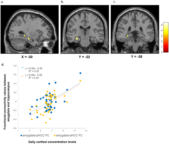
The amygdala plays a critical role in emotion. Its functional coupling with the hippocampus and ventromedial prefrontal cortex extending to a portion of the anterior cingulate cortex (ACC) is implicated in anxiogenesis and hypothalamic-pituitary-adrenal (HPA) system regulation. However, it remains unclear how amygdala-centred functional connectivity (FC) affects anxiety and cortisol concentrations in everyday life. Here, we investigate the relationship between daily cortisol concentrations (dCOR) and amygdala-centred FC during emotional processing in forty-one healthy humans. FC analyses revealed that higher dCOR predicted strengthened amygdala-centred FC with the hippocampus and cerebellum, but inhibited FC with the supramarginal gyrus and a perigenual part of the ACC (pgACC) when processing fearful faces (vs. neutral faces). Notably, the strength of amygdala-hippocampus FC mediated the positive relationship between cortisol and anxiety, specifically when the effect of amygdala-pgACC FC, a presumptive neural indicator of emotional control, was taken into account. Individuals with diminished connectivity between the amygdala and pgACC during fear-related processing might be more vulnerable to anxiogenesis as it pertains to greater circulating cortisol levels in everyday life. Individual functional patterns of amygdala-hippocampal-pgACC connectivity might provide a key to understand the complicate link between cortisol and anxiety-related behaviors.


Conflict of interest statement

Author H.T. received grants support from Astellas Pharma, Inc., MSD Inc., the Sanofi-Aventis Corp, and Otsuka Pharmaceutical Inc. All the authors except H.T. declare no conflict of interest associated with this manuscript.

Figures

Figure 1
Figure 1
Amygdala-centred functional connectivity during fearful faces processing. Seed region: the amygdala that showed significantly increased activation in response to fearful faces (vs. neutral faces) in this sample (L-Amy: MNI −21 −10 −17, 351 mm3, R-Amy: MNI 21 −10 −17, 189 mm3), which are comprised of the caudomedial portion of the amygdala. Clusters were significant at FDR-corrected p < 0.05 [both height threshold and cluster threshold (cluster-size correction)]. Abbreviations: L-Amy, left amygdala; R-Amy, right amygdala; MNI, Montreal Neurological Institute space; FDR, false-discovery rate.
Figure 2
Figure 2
The HC displays greater FC with the amygdala during the perception of fearful faces (vs. neutral faces). (a) Significantly heightened FC between the amygdala and 2 different clusters within the HC (its strength parallels dCOR levels); (b) Anterior part of the HC (MNI −30 −22 −16, 280 mm3, T = 5.41, p < 0.001); (c) Posterior part of the HC (MNI −30 −38 −4, 144 mm3, T = 4.30, p < 0.001). These clusters were also significant at FWE-corrected p < 0.05 with SVC. Colour bar indicates T-value. (d) Scatter plot of the relationship between dCOR and amygdala-HC FC values in fearful faces processing (vs. neutral faces). Amygdala-pHCC FC values (blue circle). Amygdala-aHCC FC values (orange circle). Regression line of amygdala-pHCC FC (blue dotted line). Regression line of amygdala-aHCC FC (orange dotted line). Abbreviations: HC, hippocampus; FC, functional connectivity; dCOR, daily cortisol concentrations; MNI, Montreal Neurological Institute space; SVC, small volume correction; pHCC, a posterior part of the HC; aHCC, an anterior part of the HC.
Figure 3
Figure 3
The pgACC exhibits diminished FC with the amygdala. (a) pgACC cluster found at MNI 0 30 14 (120 mm3; T  = 4.40, p < 0.001). Colour bar indicates T-value. (b) Scatter plot of the relationship between dCOR and amygdala-pgACC FC values in fearful faces processing (vs. neutral faces). Abbreviations: pgACC, perigenual anterior cingulate cortex; FC, functional connectivity; MNI, Montreal Neurological Institute space; dCOR, daily cortisol concentrations.
Figure 4
Figure 4
Results of mediation analysis. Amygdala-HC FC mediating effect on the relationship between anxiety symptoms and dCOR. The effect of the amygdala-pgACC FC was incorporated as covariate in the mediation model. Abbreviations: HC, hippocampus; FC, functional connectivity; dCOR, daily cortisol concentrations; pgACC, periginual anterior cingulate cortex. *p < 0.05, **p < 0.01, ***p < 0.001. All path coefficients are unstandardized regression weights. The value in parentheses indicates anxiety symptoms’ total effect on dCOR.
Figure 5
Figure 5
Experimental paradigm. The first display presents 4 empty frames in vertically and horizontally paired positions, with either the 2 horizontal or the 2 vertical frames being highlighted. The next display shows a central fixation point followed by paired faces and paired houses arranged in vertical or horizontal positions. The 2 highlighted frames always predict the locations of the paired houses. Both faces had either neutral or fearful expressions. The positions of the paired faces or houses (vertical or horizontal) and the facial expressions (neutral or fearful) were counterbalanced across subjects and were presented in a random order. The figure is reprinted with permission from the ref. (2010) © Macmillan Publishers Ltd., in Nature Publishing Group. The figure is a modified version of the original figure from the ref. (2001) © Cell Press.

References

    1. Reul JM, de Kloet ER. Two receptor systems for corticosterone in rat brain: microdistribution and differential occupation. Endocrinology. 1985;117:2505–11. doi: 10.1210/endo-117-6-2505. - DOI - PubMed
    1. De Kloet ER, Reul JM. Feedback action and tonic influence of corticosteroids on brain function: a concept arising from the heterogeneity of brain receptor systems. Psychoneuroendocrinology. 1987;12:83–105. doi: 10.1016/0306-4530(87)90040-0. - DOI - PubMed
    1. Herman JP, et al. Regulation of the Hypothalamic-Pituitary-Adrenocortical Stress Response. Compr Physiol. 2016;6:603–21. doi: 10.1002/cphy.c150015. - DOI - PMC - PubMed
    1. Phan K, Wager T, Taylor S, Liberzon I. Functional neuroanatomy of emotion: a meta-analysis of emotion activation studies in PET and fMRI. Neuroimage. 2002;16:331–48. doi: 10.1006/nimg.2002.1087. - DOI - PubMed
    1. Sabatinelli D, et al. Emotional perception: meta-analyses of face and natural scene processing. Neuroimage. 2011;54:2524–33. doi: 10.1016/j.neuroimage.2010.10.011. - DOI - PubMed

Publication types