Review
. 2017 Aug 3:6:1318.
doi: 10.12688/f1000research.11683.1. eCollection 2017.

Toward an understanding of the Cdc48/p97 ATPase

Affiliations
Review

Toward an understanding of the Cdc48/p97 ATPase

Nicholas Bodnar et al. F1000Res. .

Abstract

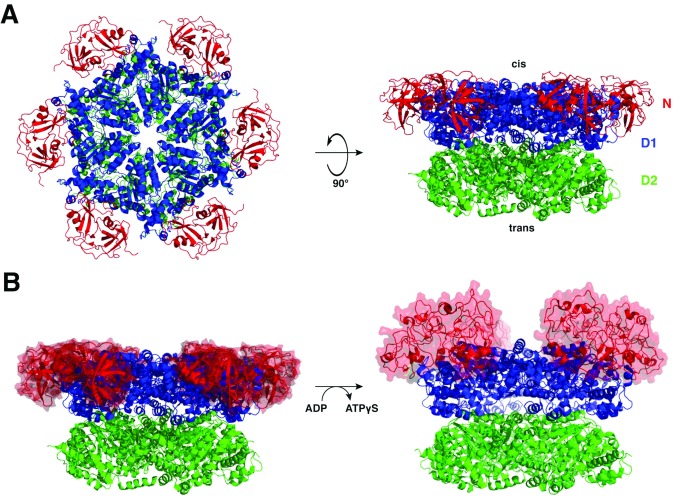
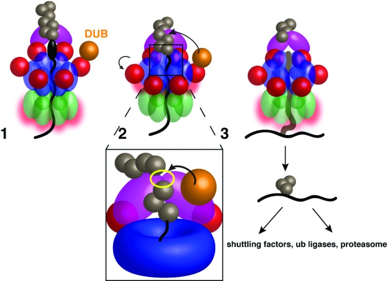
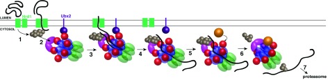
A conserved AAA+ ATPase, called Cdc48 in yeast and p97 or VCP in metazoans, plays an essential role in many cellular processes by segregating polyubiquitinated proteins from complexes or membranes. For example, in endoplasmic reticulum (ER)-associated protein degradation (ERAD), Cdc48/p97 pulls polyubiquitinated, misfolded proteins out of the ER and transfers them to the proteasome. Cdc48/p97 consists of an N-terminal domain and two ATPase domains (D1 and D2). Six Cdc48 monomers form a double-ring structure surrounding a central pore. Cdc48/p97 cooperates with a number of different cofactors, which bind either to the N-terminal domain or to the C-terminal tail. The mechanism of Cdc48/p97 action is poorly understood, despite its critical role in many cellular systems. Recent in vitro experiments using yeast Cdc48 and its heterodimeric cofactor Ufd1/Npl4 (UN) have resulted in novel mechanistic insight. After interaction of the substrate-attached polyubiquitin chain with UN, Cdc48 uses ATP hydrolysis in the D2 domain to move the polypeptide through its central pore, thereby unfolding the substrate. ATP hydrolysis in the D1 domain is involved in substrate release from the Cdc48 complex, which requires the cooperation of the ATPase with a deubiquitinase (DUB). Surprisingly, the DUB does not completely remove all ubiquitin molecules; the remaining oligoubiquitin chain is also translocated through the pore. Cdc48 action bears similarities to the translocation mechanisms employed by bacterial AAA ATPases and the eukaryotic 19S subunit of the proteasome, but differs significantly from that of a related type II ATPase, the NEM-sensitive fusion protein (NSF). Many questions about Cdc48/p97 remain unanswered, including how it handles well-folded substrate proteins, how it passes substrates to the proteasome, and how various cofactors modify substrates and regulate its function.

Keywords: ATPase; Cdc48; p97.


Conflict of interest statement

Competing interests: The authors declare that they have no competing interests.No competing interests were disclosed.No competing interests were disclosed.No competing interests were disclosed.No competing interests were disclosed.No competing interests were disclosed.

Figures

Figure 1.
Figure 1.. Structure of the Cdc48/p97 ATPase.
( A) Cdc48 is a homohexamer, and each monomer comprises an N-terminal (N) domain (red) and two AAA ATPase domains: D1 (blue) and D2 (green). The N-terminal (D1) side of the central pore is referred to as the cis side, and the C-terminal (D2) side as the trans side. ( B) ATP binding produces an upward rotation of the N domains into a so-called “up conformation”, in which they are positioned above the plane of the D1 ring. Left, ADP-bound state (PDB code 5FTK); right, ATPγS-bound state (PDB code 5FTN). PDB, Protein Data Bank.
Figure 2.
Figure 2.. Model for Cdc48 substrate unfolding.
The process begins with the N domains in the “down conformation”. In step 1, the N domains transition into the “up conformation” upon D1 ATP binding. Binding of the Ufd1/Npl4 (UN) complex to the N domains renders the complex competent for substrate binding. In step 2, the polyubiquitin chain attached to a substrate binds to the UN complex. The D1 domain stays in the ATP-bound state with the N domains in the “up conformation”. At the same time, the ATPase rate of the D2 domains is increased (red outline). In step 3, the substrate inserts into the central pore. In step 4, ATP hydrolysis by the D2 domains unfolds the substrate by pulling it through the pore to the trans side.
Figure 3.
Figure 3.. Model for substrate release from Cdc48.
Stage 1: The substrate is pulled through the double-ring ATPase, but the polyubiquitin chain is bound to the Ufd1/Npl4 (UN) complex at the cis side, preventing full substrate translocation through the pore, even with continued D2 ATP hydrolysis. A deubiquitinase (DUB) bound to the N domain cannot access the polyubiquitin chain while D1 is in the ATP-bound state and the N domains are in the “up conformation”. Stage 2: D1 hydrolyzes ATP after the substrate has initiated or completed translocation, moving the N domains into the “down conformation” and allowing DUB access to the ubiquitin chain. Inset: The likely Ub chain cleavage site is between the UN binding site (yellow outline) and the central pore; any Ub moieties proximal to the cleavage site will be translocated. Stage 3: After polyubiquitin cleavage, the oligoubiquitinated substrate proceeds fully through the pore. The ubiquitin moieties probably refold after translocation, and the substrate is transferred to downstream factors.
Figure 4.
Figure 4.. Model for Cdc48 function in endoplasmic reticulum–associated protein degradation.
In step 1, a segment of a misfolded luminal substrate is moved into the cytosol and polyubiquitinated by Hrd1. In step 2, the Cdc48 complex is bound to the cytosolic face of the endoplasmic reticulum via an interaction of its N domain with Ubx2. The complex recognizes the polyubiquitin chain via Ufd1/Npl4 (UN). In step 3, Cdc48 uses ATP hydrolysis in the D2 domains to extract the substrate from the membrane, translocating the polypeptide through its central pore. In step 4, Cdc48 completes translocation and eventually diffuses away from Ubx2, together with the bound substrate. In step 5, a deubiquitinase (DUB) such as Otu1 binds to the newly vacant N domain. In step 6, the D1 domains hydrolyze ATP, moving the N domains to the “down conformation” and allowing trimming of the ubiquitin chain. The oligoubiquitinated substrate then is released from the trans side of the pore. In step 7, the substrate is transferred to downstream factors, eventually arriving at the proteasome.

References

    1. Erzberger JP, Berger JM: Evolutionary relationships and structural mechanisms of AAA+ proteins. Annu Rev Biophys Biomol Struct. 2006;35:93–114. 10.1146/annurev.biophys.35.040405.101933 - DOI - PubMed
    1. Sauer RT, Baker TA: AAA+ proteases: ATP-fueled machines of protein destruction. Annu Rev Biochem. 2011;80:587–612. 10.1146/annurev-biochem-060408-172623 - DOI - PubMed
    1. Xia D, Tang WK, Ye Y: Structure and function of the AAA+ ATPase p97/Cdc48p. Gene. 2016;583(1):64–77. 10.1016/j.gene.2016.02.042 - DOI - PMC - PubMed
    1. Watts GD, Wymer J, Kovach MJ, et al. : Inclusion body myopathy associated with Paget disease of bone and frontotemporal dementia is caused by mutant valosin-containing protein. Nat Genet. 2004;36(4):377–81. 10.1038/ng1332 - DOI - PubMed
    1. Kimonis VE, Fulchiero E, Vesa J, et al. : VCP disease associated with myopathy, Paget disease of bone and frontotemporal dementia: review of a unique disorder. Biochim Biophys Acta. 2008;1782(12):744–8. 10.1016/j.bbadis.2008.09.003 - DOI - PubMed