. 2017 Feb;4(2):280-288.
doi: 10.1364/OPTICA.4.000280. Epub 2017 Feb 20.

Focusing light inside dynamic scattering media with millisecond digital optical phase conjugation

Affiliations

Focusing light inside dynamic scattering media with millisecond digital optical phase conjugation

Yan Liu et al. Optica. 2017 Feb.

Abstract

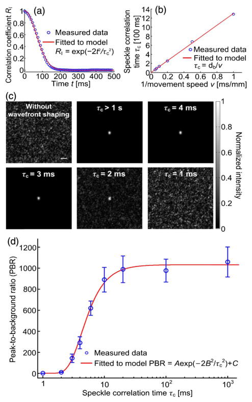
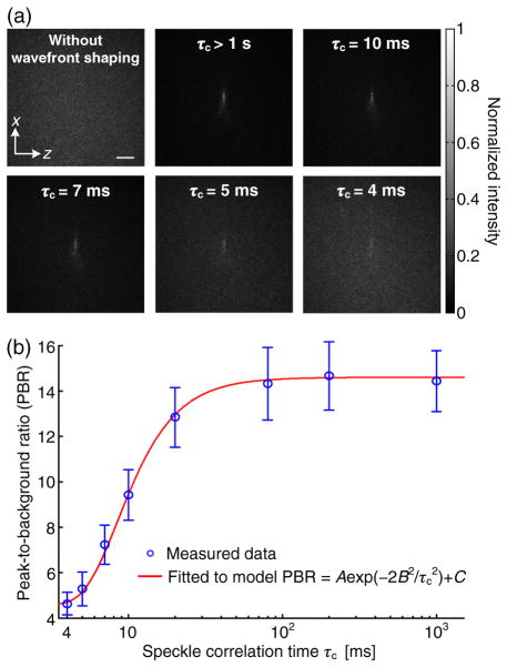
Wavefront shaping based on digital optical phase conjugation (DOPC) focuses light through or inside scattering media, but the low speed of DOPC prevents it from being applied to thick, living biological tissue. Although a fast DOPC approach was recently developed, the reported single-shot wavefront measurement method does not work when the goal is to focus light inside, instead of through, highly scattering media. Here, using a ferroelectric liquid crystal based spatial light modulator, we develop a simpler but faster DOPC system that focuses light not only through, but also inside scattering media. By controlling 2.6 × 10⁵ optical degrees of freedom, our system focused light through 3 mm thick moving chicken tissue, with a system latency of 3.0 ms. Using ultrasound-guided DOPC, along with a binary wavefront measurement method, our system focused light inside a scattering medium comprising moving tissue with a latency of 6.0 ms, which is one to two orders of magnitude shorter than those of previous digital wavefront shaping systems. Since the demonstrated speed approaches tissue decorrelation rates, this work is an important step toward in vivo deep-tissue non-invasive optical imaging, manipulation, and therapy.


Figures

Fig. 1
Fig. 1
Comparison of different wavefront modulation schemes in wavefront shaping. PBR, peak-to-background ratio.
Fig. 2
Fig. 2
DOPC using a ferroelectric liquid crystal based spatial light modulator (FLC-SLM). (a) Each FLC-SLM pixel acts as a half-wave plate. PBS, polarizing beamsplitter. (b) Optic axis orientation can be switched between two states, e1 and e2, to achieve binary-phase modulation of the incident light Ein θ = 22.5°. (c) Schematic of the setup during wavefront recording for DOPC-based light focusing through scattering media. BB, beam block; BS, beamsplitter; CL, camera lens; DOPC, digital optical phase conjugation; HWP, half-wave plate; M, mirror; MLS, motorized linear stage; MS, mechanical shutter; PC, personal computer; PCIe ×4, peripheral component interconnect express interface with four lanes; SM, scattering medium; S, sample beam; S*, phase-conjugated sample beam; and Rr and Rp, reference beams for wavefront recording and playback. The distance between SM and L6 (f = 100 mm) is 40 cm. (d) Schematic of the setup during wavefront playback for DOPC-based light focusing through scattering media. (e) Schematic of the setup for focusing light inside a scattering medium comprising two pieces of chicken tissue with ultrasound-guided DOPC. A complete schematic can be obtained by replacing the components enclosed in the dashed box in (c) and (d) with the components enclosed in the dashed box in (e). The acousto-optic modulator (AOM) is used only during wavefront recording. During wavefront playback, to verify that light is focused to the ultrasonic (US) focus, a beamsplitter (BS) reflects the focal pattern onto Camera2 (Cam2). To control the speckle correlation time on the SLM plane, a MLS moves the second piece of tissue at different speeds during the entire DOPC process (including both wavefront measurement and playback). The distance between the two pieces of tissue is 32 mm, and the distance between the ultrasonic focus and the tissue on the right side is 20 mm.
Fig. 3
Fig. 3
Workflow of TRUE optical focusing inside scattering media. A rolling shutter was used for Camera1, that is, neighboring rows are exposed successively with a 9.17 μs delay in the start times. The shutter for S (LS6, Vincent Associates) has a full-aperture transfer time of 0.8 ms, while the shutters for Rr (VSR14, Vincent Associates) and Rp (VS14, Vincent Associates) have full-aperture transfer times of 1.5 ms, because of larger aperture sizes (14 mm). FG, function generator; Ch, channel; RF, radio-frequency.
Fig. 4
Fig. 4
System performance quantification. (a) Image of the DOPC focus after light passed through an opal diffuser with a 4π scattering angle. The PBR is 5.1 × 10³. Scale bar, 100 μm. (b) Focal intensity distribution along the vertical direction.
Fig. 5
Fig. 5
Focusing light through moving scattering tissue. (a) Correlation coefficient between the speckle patterns as a function of time, when a 3 mm thick slice of chicken tissue was moved at 0.01 mm/s. Speckle correlation time τc =1.3 × 10² ms was determined for this speed. (b) Relationship between the speckle correlation time and the tissue movement speed. Errors bars are not plotted due to indiscernible lengths in the figure. (c) Images of the DOPC foci after light passed through the tissue, when the tissue was moved at different speeds. Scale bar, 100 μm. (d) PBR as a function of the speckle correlation time. The error bar shows the standard deviation of three measurements.
Fig. 6
Fig. 6
Focusing light inside a dynamic scattering medium comprised of two pieces of chicken tissue. (a) Images of the foci achieved by TRUE focusing at different speckle correlation times (τc). Scale bar, 500 μm. (b) The PBR as a function of the speckle correlation time. The error bar shows the standard deviation of three measurements.

References

    1. Ntziachristos V. Going deeper than microscopy: the optical imaging frontier in biology. Nat Methods. 2010;7:603–614. - PubMed
    1. Liu Y, Zhang C, Wang LV. Effects of light scattering on optical-resolution photoacoustic microscopy. J Biomed Opt. 2012;17:126014. - PMC - PubMed
    1. Mosk AP, Lagendijk A, Lerosey G, Fink M. Controlling waves in space and time for imaging and focusing in complex media. Nat Photonics. 2012;6:283–292.
    1. Horstmeyer R, Ruan H, Yang C. Guidestar-assisted wavefront-shaping methods for focusing light into biological tissue. Nat Photonics. 2015;9:563–571. - PMC - PubMed
    1. Vellekoop IM. Feedback-based wavefront shaping. Opt Express. 2015;23:12189–12206. - PubMed