. 2017 Jul 26;2(4):e00292-17.
doi: 10.1128/mSphere.00292-17. eCollection 2017 Jul-Aug.

Phenotypic Differences between Asian and African Lineage Zika Viruses in Human Neural Progenitor Cells

Affiliations

Phenotypic Differences between Asian and African Lineage Zika Viruses in Human Neural Progenitor Cells

Fatih Anfasa et al. mSphere. .

Abstract

Recent Zika virus (ZIKV) infections have been associated with a range of neurological complications, in particular congenital microcephaly. Human neural progenitor cells (hNPCs) are thought to play an important role in the pathogenesis of microcephaly, and experimental ZIKV infection of hNPCs has been shown to induce cell death. However, the infection efficiency and rate of cell death have varied between studies, which might be related to intrinsic differences between African and Asian lineage ZIKV strains. Therefore, we determined the replication kinetics, including infection efficiency, burst size, and ability to induce cell death, of two Asian and two African ZIKV strains. African ZIKV strains replicated to higher titers in Vero cells, human glioblastoma (U87MG) cells, human neuroblastoma (SK-N-SH) cells, and hNPCs than Asian ZIKV strains. Furthermore, infection with Asian ZIKV strains did not result in significant cell death early after infection, whereas infection with African ZIKV strains resulted in high percentages of cell death in hNPCs. The differences between African and Asian lineage ZIKV strains highlight the importance of including relevant ZIKV strains to study the pathogenesis of congenital microcephaly and caution against extrapolation of experimental data obtained using historical African ZIKV strains to the current outbreak. Finally, the fact that Asian ZIKV strains infect only a minority of cells with a relatively low burst size together with the lack of early cell death induction might contribute to its ability to cause chronic infections within the central nervous system (CNS). IMPORTANCE The mechanism by which ZIKV causes a range of neurological complications, especially congenital microcephaly, is not well understood. The fact that congenital microcephaly is associated with Asian lineage ZIKV strains raises the question of why this was not discovered earlier. One possible explanation is that Asian and African ZIKV strains differ in their abilities to infect cells of the CNS and to cause neurodevelopmental problems. Here, we show that Asian ZIKV strains infect and induce cell death in human neural progenitor cells-which are important target cells in the development of congenital microcephaly-less efficiently than African ZIKV strains. These features of Asian ZIKV strains likely contribute to their ability to cause chronic infections, often observed in congenital microcephaly cases. It is therefore likely that phenotypic differences between ZIKV strains could be, at least in part, responsible for the ability of Asian ZIKV strains to cause congenital microcephaly.

Keywords: African strains; Asian strains; Zika virus; cell death; growth kinetics; human neural progenitor cells; neuronal cells; one-step growth curve; pathogenesis; phenotype.


Figures
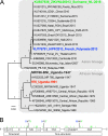
FIG 1
FIG 1
Phylogenetic analysis of ZIKV strains used in this study and genomic organization and mutations between the Asian lineage ZIKV strains. (A) Nucleotide sequences of representative Zika virus genomes were analyzed, and a phylogenetic tree was constructed using the PhyML method. Values at branches show the result of the approximate likelihood ratio; values of <0.70 are not shown. (B) Genome organization and mutations between Asian lineage H/PF/2013 (ZIKVAS-FP13) and ZIKVNL00013 (ZIKVAS-Sur16) ZIKV strains.
FIG 2
FIG 2
Growth curves of ZIKV strains on Vero, SK-N-SH, and U87-MG cells and hNPCs. (A and B) Growth curves of Asian lineage strains H/PF/2013 (ZIKVAS-FP13 [blue lines]) and ZIKVNL00013 (ZIKVAS-Sur16 [green lines]) and African lineage MR766 (ZIKVAF-MR766 [black lines]) and 976 Uganda (ZIKVAF-976 [red lines]) on Vero, human neuroblastoma (SK-N-SH), and human glioblastoma (U87-MG) cells and human neuronal progenitor cells (hNPCs) at MOI of 0.1 (A) and 0.01 (B). Data are presented as means with standard deviations from at least 3 independent experiments. Statistical significance was calculated using the Student t test in comparison with ZIKVAF-MR766. *, P ≤ 0.05; **, P ≤ 0.01; ***, P ≤ 0.001; ****, P ≤ 0.0001. TCID50, 50% tissue culture infectious dose.
FIG 3
FIG 3
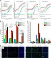
One-step growth curve (OSGC) kinetics of Asian and African lineage ZIKV strains. (A) OSGCs of Asian lineage strains H/PF/2013 (ZIKVAS-FP13 [blue lines]) and ZIKVNL00013 (ZIKVAS-Sur16 [green lines]) and African lineage MR766 (ZIKVAF-MR766 [black lines]) and 976 Uganda (ZIKVAF-976 [red lines]) on Vero, human neuroblastoma (SK-N-SH), and human glioblastoma (U87-MG) cells and human neuronal progenitor cells (hNPCs). (B) Percentage of ZIKV infection determined by immunofluorescent microscopy of two Asian and two African ZIKV strains. (C) Number of infectious viruses produced per cell (burst size) for each virus in the 4 different cell lines. (D) Representative immunofluorescent images of ZIKV-infected cells stained for ZIKV antigen (green). Magnification, ×200. For panels A and B, data are presented as means with standard deviations and nonlinear curve fit for at least 3 independent experiments. For panel C, data are presented as means with standard errors of the means from at least 3 independent experiments. Statistical significance was calculated using a one-way ANOVA with Tukey’s multiple comparisons test for panel A. For panels B and C, the Student t test was used. *, P ≤ 0.05; **, P ≤ 0.01; ***, P ≤ 0.001; ****, P ≤ 0.0001. TCID50, 50% tissue culture infectious dose.
FIG 4
FIG 4
Ability to cause cell death of African and Asian lineage ZIKV strains in human neural progenitor cells. (A) Percentage of human neural progenitor cells infected with African lineage ZIKV strain ZIKVAF-MR766 (black lines) and Asian lineage ZIKV strains H/PF/2013 (ZIKVAS-FP13 [blue lines]) and ZIKVNL00013 (ZIKVAS-Sur16 [green lines]) and percentage of TUNEL-positive cells measured over 72 h. The left y axis represents the percentage of cells infected with ZIKV, and the right y axis represents the percentage of TUNEL-positive cells. Data are presented as means with standard errors of the means from at least 3 independent experiments. (B) Representative immunofluorescent images of human neural progenitor cells infected with different ZIKV strains 48 h postinfection and double stained for ZIKV antigen (red) and TUNEL (green). Asterisks indicate double-positive cells. Magnification, ×200.

Similar articles

Cited by

References

    1. Ritter JM, Martines RB, Zaki SR. 2017. Zika virus: pathology from the pandemic. Arch Pathol Lab Med 141:49–59. doi:10.5858/arpa.2016-0397-SA. - DOI - PubMed
    1. Li H, Saucedo-Cuevas L, Shresta S, Gleeson JG. 2016. The neurobiology of Zika virus. Neuron 92:949–958. doi:10.1016/j.neuron.2016.11.031. - DOI - PubMed
    1. Weaver SC. 2017. Emergence of epidemic Zika virus transmission and congenital Zika syndrome: are recently evolved traits to blame? mBio 8:e02063-16. doi:10.1128/mBio.02063-16. - DOI - PMC - PubMed
    1. Wang L, Valderramos SG, Wu A, Ouyang S, Li C, Brasil P, Bonaldo M, Coates T, Nielsen-Saines K, Jiang T, Aliyari R, Cheng G. 2016. From mosquitos to humans: genetic evolution of Zika virus. Cell Host Microbe 19:561–565. doi:10.1016/j.chom.2016.04.006. - DOI - PMC - PubMed
    1. Pettersson JH, Eldholm V, Seligman SJ, Lundkvist Å, Falconar AK, Gaunt MW, Musso D, Nougairède A, Charrel R, Gould EA, de Lamballerie X. 2016. How did Zika virus emerge in the Pacific Islands and Latin America? mBio 7:e01239-16. doi:10.1128/mBio.01239-16. - DOI - PMC - PubMed