. 2018 Jun;55(6):5047-5058.
doi: 10.1007/s12035-017-0705-1. Epub 2017 Aug 16.

Inhibition of γ-Secretase Leads to an Increase in Presenilin-1

Affiliations

Inhibition of γ-Secretase Leads to an Increase in Presenilin-1

Aitana Sogorb-Esteve et al. Mol Neurobiol. 2018 Jun.

Abstract

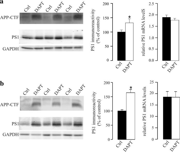
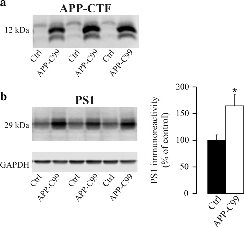
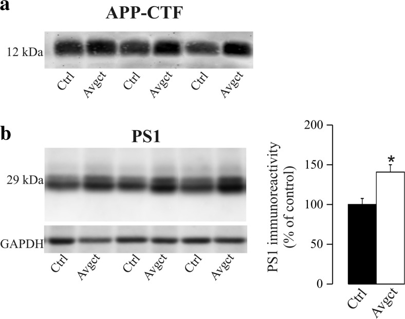
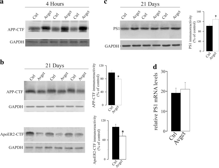
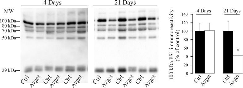
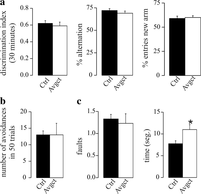
γ-Secretase inhibitors (GSIs) are potential therapeutic agents for Alzheimer's disease (AD); however, trials have proven disappointing. We addressed the possibility that γ-secretase inhibition can provoke a rebound effect, elevating the levels of the catalytic γ-secretase subunit, presenilin-1 (PS1). Acute treatment of SH-SY5Y cells with the GSI LY-374973 (N-[N-(3,5-difluorophenacetyl)-L-alanyl]-S-phenylglycine t-butyl ester, DAPT) augments PS1, in parallel with increases in other γ-secretase subunits nicastrin, presenilin enhancer 2, and anterior pharynx-defective 1, yet with no increase in messenger RNA expression. Over-expression of the C-terminal fragment (CTF) of APP, C99, also triggered an increase in PS1. Similar increases in PS1 were evident in primary neurons treated repeatedly (4 days) with DAPT or with the GSI BMS-708163 (avagacestat). Likewise, rats examined after 21 days administered with avagacestat (40 mg/kg/day) had more brain PS1. Sustained γ-secretase inhibition did not exert a long-term effect on PS1 activity, evident through the decrease in CTFs of APP and ApoER2. Prolonged avagacestat treatment of rats produced a subtle impairment in anxiety-like behavior. The rebound increase in PS1 in response to GSIs must be taken into consideration for future drug development.

Keywords: Alzheimer’s disease; Presenilin-1; Therapy; γ-Secretase inhibitor.


Conflict of interest statement

Conflict of Interest

The authors declare that they have no competing interests.

Figures

Fig. 1
Fig. 1
GSI DAPT treatment augments PS1 in SH-SY5Y cells and in mouse primary neurons. a SH-SY5Y cells were treated for 18 h (acutely) with DAPT (5 μM) or the vehicle alone (control; Ctrl). Cell extracts were probed with antibody C1-6.1, against the APP C-terminal, to demonstrate the accumulation of the APP-CTF in treated cells as a result of the inhibition of γ-secretase processing. Cell extracts were also probed for PS1 with an anti-N-terminal antibody. Equivalent amounts of protein were loaded in each lane, and GAPDH was used as a loading control. Representative blots and densitometric quantification of the immunoreactivity are shown. Relative expression of PS1 mRNA was also analyzed by qRT-PCR. Transcript levels were calculated by the comparative 2−ΔCt method with respect to GAPDH cDNA. b Primary neurons were treated with DAPT (2 μM) or the vehicle alone (Ctrl) for four consecutive days. Cell extracts were probed for APP-CTF and PS1 and for GAPDH as a loading control. The densitometric quantification for PS1-NTF is shown, as well the relative mRNA levels of the PS1 transcript. The data represent the means ± SEM of at least n = 10 independent determinations (obtained from two independent sets of experiments): *p < 0.05
Fig. 2
Fig. 2
Effects of the modulation of APP-CTF expression on PS1 levels. SH-SY5Y cells were transfected with APP-C99 cDNA, the β-secretase-derived CTF of APP, or with a control vector (Ctrl). a Immunodetection of APP-CTF with the anti-APP C-terminal antibody C1-6.1 served to assess the efficiency of over-expression. The identity of the increased immunoreactive band was also tested with the 6E10 antibody, which recognizes an epitope present in the N-terminal of APP-C99 (not shown). b The immunodetection and densitometric quantification of PS1 immunoreactivity in transfected cells are shown. The data are presented relative to control cells, expressed as the means ± SEM of at least 12 independent determinations (obtained from two independent sets of experiments): *p = 0.007
Fig. 3
Fig. 3
Increased PS1 levels in neurons treated with the GSI, avagacestat. Primary neurons were treated with avagacestat (2 μM, Avgct) or the vehicle alone (Ctrl), and the cell extracts were probed for a APP-CTF and b PS1. Representative blots and their densitometric quantification are shown. The data presented are relative to the Ctrl cells, expressed as the means ± SEM of at least ten independent determinations (obtained from two independent sets of experiments): *p = 0.007
Fig. 4
Fig. 4
Effect of prolonged inhibition of γ-secretase by avagacestat on γ-secretase substrates and PS1 in the cortex of rats treated for 21 days. Rats were treated daily with the GSI avagacestat (40 mg/kg, Avgct) or the vehicle alone (control; Ctrl) for 21 days, and they were sacrificed 4 h after the last dose. a As a control of the effective γ-secretase inhibition by the GSI in the brain, APP-CTF levels (probed with antibody C1-6.1) were firstly evaluated in rats sacrificed 4 h after a single dose of avagacestat (n = 6 per group). b The levels of APP-CTF and ApoER2-CTF were estimated in rats treated with avagacestat for 21 days; representative blots and densitometric quantifications are shown. c PS1 levels were also evaluated in Western blots of the same brain hemi-cortex extracts. GAPDH was used as a loading control. d Relative PS1 mRNA was analyzed by qRT-PCR in the other rat hemi-cortices obtained after 21 days of treatment (n = 10 per group). The data are presented relative to the control rats and expressed as the means ± SEM (n = 10 per group): * p < 0.05 significantly different from the controls
Fig. 5
Fig. 5
Effect of avagacestat on PS1 levels in CSF of rats treated for 4 and 21 days. Rats were administered avagacestat (40 mg/kg, Avgct) or the vehicle alone (Ctrl) daily over 4 or 21 days. Soluble PS1 complexes were also evaluated in Western blots of CSF samples from Avgct-treated and control rats (n = 7 per group). CSF-PS1 complexes were detected with and N-terminal antibody, which predominantly recognized stable complexes of approximately 100 kDa, together with less abundant 80-, 70-, and 50-kDa complexes, as well monomers of 29 kDa. Previous studies indicated that these CSF-PS1 complexes represent aggregates of PS1-NTF and CTF [40, 41]. The densitometric quantification of the major CSF-PS1 100-kDa complex is shown. The data are presented relative to the control rats, expressed as the means ± SEM: *p < 0.05
Fig. 6
Fig. 6
Results of the behavioral tests in rats treated 21 days with avagacestat. a Novel spatial recognition memory in the Y-maze in rats treated with avagacestat for 21 days (Avgct) and in the vehicle-treated controls (Ctrl). The time spent in each arm was recorded in order to calculate the discrimination index after a 30-min inter-trial interval. b Result of the active avoidance test documenting the number of attempts made to avoid the foot shock. c Beam walking test in which the number of slips and the latency to cross were scored. The values are the means ± SEM (n = 10 for each group): *p < 0.05

Similar articles

Cited by

References

    1. Scheltens P, Blennow K, Breteler MM, De Strooper B, Frisoni GB, Salloway S, et al. Alzheimer’s disease. Lancet. 2016;388:505–517. doi: 10.1016/S0140-6736(15)01124-1. - DOI - PubMed
    1. Hardy J, Selkoe DJ. The amyloid hypothesis of Alzheimer’s disease: progress and problems on the road to therapeutics. Science. 2002;297:353–356. doi: 10.1126/science.1072994. - DOI - PubMed
    1. Schneider LS, Mangialasche F, Andreasen N, Feldman H, Giacobini E, Jones R, et al. Clinical trials and late-stage drug development for Alzheimer’s disease: an appraisal from 1984 to 2014. J Intern Med. 2014;275:251–283. doi: 10.1111/joim.12191. - DOI - PMC - PubMed
    1. Portelius E, Price E, Brinkmalm G, Stiteler M, Olsson M, Persson R, et al. A novel pathway for amyloid precursor protein processing. Neurobiol Aging. 2011;32:1090–1098. doi: 10.1016/j.neurobiolaging.2009.06.002. - DOI - PubMed
    1. Haass C, Kaether C, Thinakaran G, Sisodia S. Trafficking and proteolytic processing of APP. Cold Spring Harb Perspect Med. 2012;2:a006270. doi: 10.1101/cshperspect.a006270. - DOI - PMC - PubMed