Case Reports
. 2018 Apr;20(4):411-419.
doi: 10.1038/gim.2017.115. Epub 2017 Aug 17.

Monoallelic and biallelic CREB3L1 variant causes mild and severe osteogenesis imperfecta, respectively

Affiliations
Case Reports

Monoallelic and biallelic CREB3L1 variant causes mild and severe osteogenesis imperfecta, respectively

Rachel B Keller et al. Genet Med. 2018 Apr.

Abstract

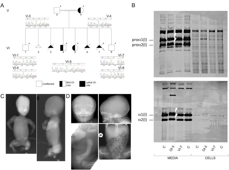
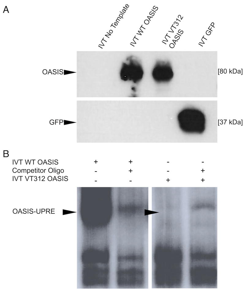
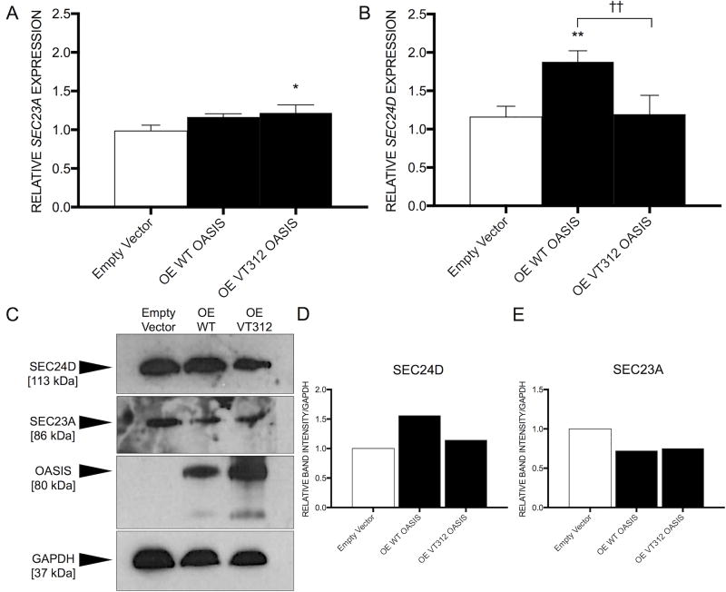
PurposeOsteogenesis imperfecta (OI) is a heritable skeletal dysplasia. Dominant pathogenic variants in COL1A1 and COL1A2 explain the majority of OI cases. At least 15 additional genes have been identified, but those still do not account for all OI phenotypes that present. We sought the genetic cause of mild and lethal OI phenotypes in an unsolved family.MethodsWe performed exome sequencing on seven members of the family, both affected and unaffected.ResultsWe identified a variant in cyclic AMP responsive element binding protein 3-like 1 (CREB3L1) in a consanguineous family. The variant caused a prenatal/perinatal lethal OI in homozygotes, similar to that seen in OI type II as a result of mutations in type I collagen genes, and a mild phenotype (fractures, blue sclerae) in multiple heterozygous family members. CREB3L1 encodes old astrocyte specifically induced substance (OASIS), an endoplasmic reticulum stress transducer. The variant disrupts a DNA-binding site and prevents OASIS from acting on its transcriptional targets including SEC24D, which encodes a component of the coat protein II complex.ConclusionThis report confirms that CREB3L1 is an OI-related gene and suggests the pathogenic mechanism of CREB3L1-associated OI involves the altered regulation of proteins involved in cellular secretion.


Conflict of interest statement

CONFLICT OF INTEREST The authors declare no conflict of interest.

Figures

Figure 1
Figure 1
Homozygosity and heterozygosity for a 3bp deletion in CREB3L1 that results in a single amino acid deletion in OASIS (c.934_936delAAG [p.Lys312del]) are consistent with the variable severity of OI phenotypes. A) Pedigree of the CREB3L1 OI family. Arrow indicates the proband. Asterisks denote the family members whose exomes were sequenced. Chromatograms from Sanger sequence analysis confirming the exome findings are shown. B) Effect of the CREB3L1 variant on type I collagen biochemistry. Collagen alpha chains synthesized by skin fibroblasts from the proband (VI-7) are structurally equivalent to those from control fibroblasts (C), in contrast to those from a typical OI type II patient (OI II) with a COL1A1 glycine substitution (p.Gly818Cys), in which overmodification results in both delayed procollagen mobility (upper panel) (arrows) and delayed α1(I) and α2(I) chain mobility when procollagens are digested with pepsin (lower panel) (arrows). We do not see abnormal retention of procollagens nor of alpha chains in the proband fibroblasts; as skin fibroblasts do not express CREB3L1, we would not expect OASIS to have a role in regulating secretion in these particular cells. C) Anterior-posterior (left) and lateral (right) radiographs from VI-3 (homozygous for the 3bp deletion in CREB3L1) following 18 weeks gestation termination. There is virtually no calvarial mineralization. Bones in all visible extremities are telescoped and very irregular. The ribs are thin and wavy and there is mild platyspondyly. D) Radiographs of VI-4 (heterozygous for the 3bp deletion in CREB3L1) at birth. The calvarium is thin and the anterior and posterior fontanelles are large, but there are no Wormian bones. The ribs are thin and there is mild platyspondyly. There is marked bowing of the right femur.
Figure 2
Figure 2
Old Astrocyte Specifically-Induced Substance (OASIS). OASIS is a 519aa protein with a basic leucine zipper (bZIP) domain common to other CREB/ATF family members and a transmembrane domain (TM) that anchors it in the RER membrane. When ER stress challenges the cell, the full-length OASIS protein is freed from the ER membrane and is cleaved through regulated intramembrane proteolysis (RIP) at the S1P and S2P sites. The conserved DNA binding domain is shown, with the deleted lysine residue in bold. Italics above show an overlapping predicted bipartite nuclear localization sequence (NLS). The deleted lysine residue is conserved in vertebrates (GERP=4.69; PhastCons=1).
Figure 3
Figure 3
Effect Of The Variant On OASIS Function. A) IVT. The variant protein (VT312) is stably expressed and does not appear truncated compared to wild type (WT) protein. IVT with no DNA input or with a construct bearing GFP DNA were included as (−) and (+) controls for in vitro transcription, respectively. Western Blot. B) The family variant causes a defect in DNA binding. A shift is seen when IVT WT OASIS is incubated with the radiolabeled oligo containing the UPRE-like sequence (lane 1). This shift is competed away when unlabeled oligo is added (lane 2). The shift is absent when radiolabeled oligo is incubated with IVT VT312 OASIS (lane 3). Background bands are likely binding interactions between labeled oligo and proteins from the HeLa lysate used to perform IVT. EMSA.
Figure 4
Figure 4
OASIS regulates expression of SEC24D, but not SEC23A. A) SEC23A, encoding a component of the COPII complex, is not significantly upregulated in the presence of WT OASIS under normal conditions. Apparently increased expression in the presence of VT312 OASIS is significant (P=0.03), but given that there is no significant difference in SEC23A expression between WT- and VT312-overexpressing cells, we conclude that SEC23A is not regulated by OASIS. *P <0.05 versus empty vector-transfected control (Tukey Test). RT-qPCR. B) A gene encoding another component of the COPII complex, SEC24D, is significantly upregulated in the presence of WT OASIS (P=0.007 versus empty vector; P=0.009 versus VT312-transfected), but not in the presence of non-functional VT312 OASIS. **P <0.01 versus empty vector-transfected control. ††P <0.01 versus the indicated group (Tukey Test). Y-axes represent relative mRNA expression normalized by the level of GAPDH. Values shown are the mean of three independent experiments (n=3). Error bars, S.D. RT-qPCR. C) Immunoblotting of whole cell lysates from HEK293 overexpressing either WT or VT312 OASIS is consistent with the expression studies, showing that D) the amount of SEC24D protein was augmented upon WT OASIS overexpression, while E) SEC23A protein levels were static.

References

    1. Rauch F, Glorieux FH. Osteogenesis Imperfecta. The Lancet. 2004;363(9418):1377–85. doi: 10.1016/s0140-6736(04)16051-0. - DOI - PubMed
    1. Byers P, Pyott S. Recessively inherited forms of osteogenesis imperfecta. Annu Rev Genet. 2012;46:475–97. doi: 10.1146/annurev-genet-110711-155608. - DOI - PubMed
    1. Harrington J, Sochett E, Howard A. Update on the evaluation and treatment of osteogenesis imperfecta. Pediatr Clin N Am. 2014;61(6):1243–57. doi: 10.1016/j.pcl.2014.08.010. - DOI - PubMed
    1. Semler O, Garbes L, Keupp K, et al. A mutation in the 5'-UTR of IFITM5 creates an in-frame start codon and causes autosomal-dominant osteogenesis imperfecta type V with hyperplastic callus. Am J Hum Genet. 2012;91(2):349–57. doi: 10.1016/j.ajhg.2012.06.011. - DOI - PMC - PubMed
    1. Guillen-Navarro E, Ballesta-Martinez MJ, Valencia M, et al. Two mutations in IFITM5 causing distinct forms of osteogenesis imperfecta. Am J Med Genet A. 2014;164A(5):1136–42. doi: 10.1002/ajmg.a.36409. - DOI - PubMed

Publication types

MeSH terms

Substances