. 2017 Oct;41(10):1381-1389.
doi: 10.1097/PAS.0000000000000900.

Cutaneous Eruptions in Patients Receiving Immune Checkpoint Blockade: Clinicopathologic Analysis of the Nonlichenoid Histologic Pattern

Affiliations

Cutaneous Eruptions in Patients Receiving Immune Checkpoint Blockade: Clinicopathologic Analysis of the Nonlichenoid Histologic Pattern

Genevieve J Kaunitz et al. Am J Surg Pathol. 2017 Oct.

Abstract

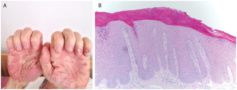
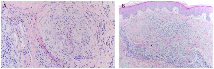
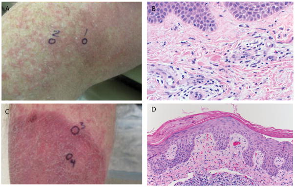
Cutaneous eruptions are among the most common immune-related adverse events (irAEs) associated with anti-programmed cell death protein 1/programmed cell death ligand 1 therapy, and are often clinically and histologically characterized as lichenoid. Nonlichenoid patterns may also occur and are likely to be encountered by surgical pathologists, given the increasing clinical use of these agents. The purpose of this study is to describe the histopathologic features of nonlichenoid cutaneous irAEs from patients receiving anti-programmed cell death protein 1/programmed cell death ligand 1 therapies for a variety of underlying advanced malignancies. Sixteen patients with 17 biopsied eruptions were included from 2 academic institutions with extensive experience administering and monitoring responses to immune checkpoint blockade as well as treating the potential side effects. Eruptions occurred a median of 10 days (range, 1 d to 11.4 mo) after treatment initiation. Nearly half of specimens demonstrated either a psoriasiform/spongiotic or an urticarial-type reaction pattern on histologic review. Patterns consistent with Grover disease, bullous pemphigoid, and granulomatous dermatitis were also observed. Nearly two-thirds of patients required systemic corticosteroids for treatment of the cutaneous irAE, and 19% of patients discontinued immunotherapy due to their skin eruptions. 75% of patients showed an objective antitumor response. The diverse array of nonlichenoid cutaneous irAE presented here should reflect and inform the scope of histologic patterns encountered by the practicing surgical pathologist. Such eruptions are seen in patients with a variety of underlying tumor types, many of whom ultimately demonstrate a favorable response to immune checkpoint blockade.


Conflict of interest statement

Conflict of interest disclosures: JMT has received research support from Bristol-Myers Squibb and is a consultant for Bristol-Myers Squibb, Merck, and AstraZeneca. JRB has received funding from Bristol-Myers Squibb and is an advisory board member (uncompensated). DTL has received research support from Merck, Bristol-Myers Squibb, and Aduro Biotech and speaking honoraria from Merck, and is a consultant for Merck. LAD is a founder of PapGene, Inc., Personal Genome Diagnostics (PGDx) and PagerBox and a consultant to Merck, Illumina, PGDx and Cell Design Labs. The first two of these companies, as well as other companies, have licensed technologies from Johns Hopkins University, on which LAD is an inventor. These licenses and relationships are associated with equity or royalty payments to LAD. The terms of these arrangements are being managed by Johns Hopkins University in accordance with its conflict of interest policies. CGD has received research funding from Bristol-Myers Squibb (IIoN grant), has consulted for Merck, Genentech, and MedImmune (AstraZeneca), and has licensed patents to MedImmune (AstraZeneca), Bristol-Myers Squibb, and Potenza Therapeutics. EJL has received research support from Genentech and is a consultant for Bristol-Myers Squibb, EMD Serono, Merck, and Novartis. MDH has received research funding from Bristol-Myers Squibb and Genentech, and has received consulting fees from Merck, Genentech, Bristol-Myers Squibb, AstraZeneca, Neon, and Inovio. For the remaining authors none were declared.

Figures

Figure 1
Figure 1
Patient 1. (A) Well-demarcated scaly plaques with 1–2 mm isolated vesicles on the bilateral palms; (B) Psoriasiform epidermal acanthosis and hypogranulosis with overlying parakeratotic scale containing neutrophils (100× original magnification).
Figure 2
Figure 2
Patient 10. (A) Multiple erythematous discrete 2–3 mm urticarial papules on distal forearm; (B) Focal acantholytic dyskeratosis, consistent with a Grover’s-like eruption (100×, original magnification).
Figure 3
Figure 3
Two patients (15 and 16) with underlying NSCLC developed granulomatous dermatitis after receiving combination anti-PD-1 and anti-CTLA-4. Histologic patterns were of (A) sarcoidal-type granulomas and (B) a granuloma-annulare pattern (200× and 100× original magnification, respectively).
Figure 4
Figure 4
Patient 4. (A) Two tense bullae (white arrows) on urticarial base adjacent to ostomy site on lower abdomen; (B) Subepidermal bullous dermatosis with prominent eosinophils, consistent with bullous pemphigoid (200×, original magnification); (C) Pre-treatment computed tomographic (CT) scan showing 12 × 6 cm left pelvic tumor mass (early tumor regression was observed at the time of first post-treatment CT scan, 2 weeks after initiation of treatment); (D) An additional follow-up CT scan image 16.5 months after treatment initiation and approximately 5 months after bullous pemphigoid eruption, showing marked tumor regression (4.6 × 1.3 cm).
Figure 5
Figure 5
Patient 12. (A) Urticarial plaques on medial thigh; (B) Superficial perivascular dermatitis including scattered eosinophils and rare neutrophils (400× original magnification); (C) Psoriasiform plaque on lower leg; (D) Psoriasiform and spongiotic dermatitis (200× original magnification). All photomicrographs are of skin biopsy specimens stained with hematoxylin and eosin (H&E).

References

    1. Topalian SL, Hodi FS, Brahmer JR, et al. Safety, activity, and immune correlates of anti-PD-1 antibody in cancer. N Engl J Med. 2012;366(26):2443–2454. - PMC - PubMed
    1. Brahmer JR, Tykodi SS, Chow LQ, et al. Safety and activity of anti-PD-L1 antibody in patients with advanced cancer. N Engl J Med. 2012;366(26):2455–2465. - PMC - PubMed
    1. Freeman-Keller M, Kim Y, Cronin H, Richards A, Gibney G, Weber JS. Nivolumab in Resected and Unresectable Metastatic Melanoma: Characteristics of Immune-Related Adverse Events and Association with Outcomes. Clin Cancer Res. 2016;22(4):886–894. - PMC - PubMed
    1. Eigentler TK, Hassel JC, Berking C, et al. Diagnosis, monitoring and management of immune-related adverse drug reactions of anti-PD-1 antibody therapy. Cancer Treat Rev. 2016;45:7–18. - PubMed
    1. Motzer RJ, Escudier B, McDermott DF, et al. Nivolumab versus Everolimus in Advanced Renal-Cell Carcinoma. N Engl J Med. 2015;373(19):1803–1813. - PMC - PubMed