

Diffusion tensor imaging profiles reveal specific neural tract distortion in normal pressure hydrocephalus
- PMID: 28817574
- PMCID: PMC5560677
- DOI: 10.1371/journal.pone.0181624
Diffusion tensor imaging profiles reveal specific neural tract distortion in normal pressure hydrocephalus
Abstract
Background: The pathogenesis of normal pressure hydrocephalus (NPH) remains unclear which limits both early diagnosis and prognostication. The responsiveness to intervention of differing, complex and concurrent injury patterns on imaging have not been well-characterized. We used diffusion tensor imaging (DTI) to explore the topography and reversibility of white matter injury in NPH pre- and early after shunting.
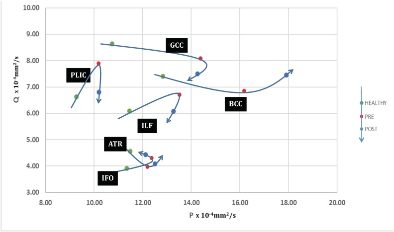
Methods: Twenty-five participants (sixteen NPH patients and nine healthy controls) underwent DTI, pre-operatively and at two weeks post-intervention in patients. We interrogated 40 datasets to generate a full panel of DTI measures and corroborated findings with plots of isotropy (p) vs. anisotropy (q).
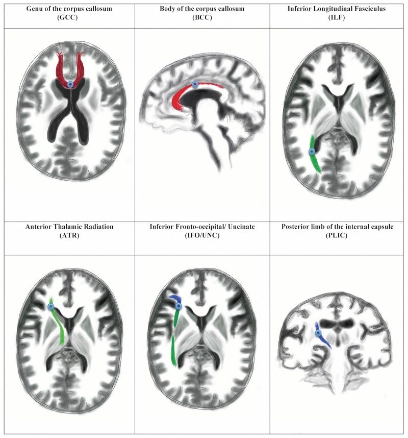
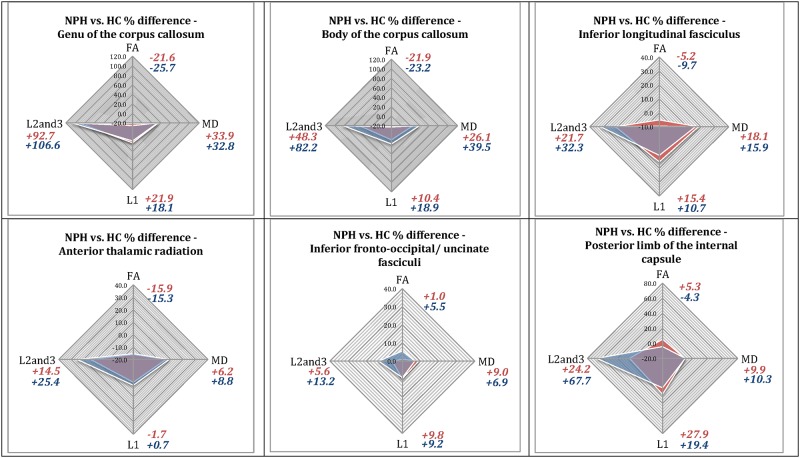
Results: Concurrent examination of DTI measures revealed distinct profiles for NPH patients vs. controls. PQ plots demonstrated that patterns of injury occupied discrete white matter districts. DTI profiles for different white matter tracts showed changes consistent with i) predominant transependymal diffusion with stretch/ compression, ii) oedema with or without stretch/ compression and iii) predominant stretch/ compression. Findings were specific to individual tracts and dependent upon their proximity to the ventricles. At two weeks post-intervention, there was a 6·7% drop in axial diffusivity (p = 0·022) in the posterior limb of the internal capsule, compatible with improvement in stretch/ compression, that preceded any discernible changes in clinical outcome. On PQ plots, the trajectories of the posterior limb of the internal capsule and inferior longitudinal fasciculus suggested attempted 'round trips'. i.e. return to normality.
Conclusion: DTI profiling with p:q correlation may offer a non-invasive biomarker of the characteristics of potentially reversible white matter injury.
Conflict of interest statement
Figures
References
-
- Adams RD, Fisher CM, Hakim S, Ojemann RG, Sweet WH. Symptomatic Occult Hydrocephalus with "Normal" Cerebrospinal-Fluid Pressure. A Treatable Syndrome. The New England journal of medicine. 1965;273:117–26. doi: 10.1056/NEJM196507152730301 . - DOI - PubMed
-
- Juss J, Keong N, Forsyth D, Pickard J. Normal pressure hydrocephalus. CME Journal Geriatric Medicine. 2008;10(2):62–7.
-
- Factora R. When do common symptoms indicate normal pressure hydrocephalus? Cleveland Clinic journal of medicine. 2006;73(5):447–50, 52,, 55–6 passim. . - PubMed
-
- Bech RA, Juhler M, Waldemar G, Klinken L, Gjerris F. Frontal brain and leptomeningeal biopsy specimens correlated with cerebrospinal fluid outflow resistance and B-wave activity in patients suspected of normal-pressure hydrocephalus. Neurosurgery. 1997;40(3):497–502. . - PubMed
-
- Momjian S, Owler BK, Czosnyka Z, Czosnyka M, Pena A, Pickard JD. Pattern of white matter regional cerebral blood flow and autoregulation in normal pressure hydrocephalus. Brain: a journal of neurology. 2004;127(Pt 5):965–72. doi: 10.1093/brain/awh131 . - DOI - PubMed
MeSH terms
Grants and funding
LinkOut - more resources
Full Text Sources
Other Literature Sources
Medical
