

Corneal confocal microscopy is a rapid reproducible ophthalmic technique for quantifying corneal nerve abnormalities
- PMID: 28817609
- PMCID: PMC5560560
- DOI: 10.1371/journal.pone.0183040
Corneal confocal microscopy is a rapid reproducible ophthalmic technique for quantifying corneal nerve abnormalities
Abstract
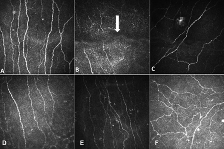
Purpose: To assess the effect of applying a protocol for image selection and the number of images required for adequate quantification of corneal nerve pathology using in vivo corneal confocal microscopy (IVCCM).
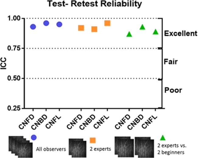
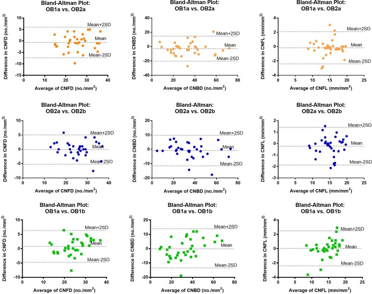
Methods: IVCCM was performed in 35 participants by a single examiner. For each participant, 4 observers used a standardized protocol to select 6 central corneal nerve images to assess the inter-observer variability. Furthermore, images were selected by a single observer on two occasions to assess intra-observer variability and the effect of sample size was assessed by comparing 6 with 12 images. Corneal nerve fiber density (CNFD), branch density (CNBD) and length (CNFL) were quantified using fully automated software. The data were compared using the intra class correlation coefficient (ICC) and Bland-Altman agreement plots for all experiments.
Results: The ICC values for CNFD, CNBD and CNFL were 0.93 (P<0.0001), 0.96 (P<0.0001) and 0.95 (P<0.0001) for inter-observer variability and 0.95 (P<0.0001), 0.97 (P<0.001) and 0.97 (P<0.0001) for intra-observer variability. For sample size variability, ICC values were 0.94 (P<0.0001), 0.95 (P<0.0001), and 0.96 (P<0.0001) for CNFD, CNBD and CNFL. Bland-Altman plots showed excellent agreement for all parameters.
Conclusions: This study shows that implementing a standardized protocol to select IVCCM images results in high intra and inter-observer reproducibility for all corneal nerve parameters and 6 images are adequate for analysis. IVCCM could therefore be deployed in large multicenter clinical trials with confidence.
Conflict of interest statement
Figures
References
-
- Petropoulos IN, Green P, Chan AW, Alam U, Fadavi H, Marshall A, et al. Corneal confocal microscopy detects neuropathy in patients with type 1 diabetes without retinopathy or microalbuminuria. PloS one. 2015;10(4):e0123517 Epub 2015/04/09. doi: 10.1371/journal.pone.0123517 ; PubMed Central PMCID: PMC4390357. - DOI - PMC - PubMed
-
- Kass-Iliyya L, Javed S, Gosal D, Kobylecki C, Marshall A, Petropoulos IN, et al. Small fiber neuropathy in Parkinson's disease: A clinical, pathological and corneal confocal microscopy study. Parkinsonism & related disorders. 2015;21(12):1454–60. doi: 10.1016/j.parkreldis.2015.10.019 ; PubMed Central PMCID: PMC4671992. - DOI - PMC - PubMed
-
- Ferdousi M, Azmi S, Petropoulos IN, Fadavi H, Ponirakis G, Marshall A, et al. Corneal Confocal Microscopy Detects Small Fibre Neuropathy in Patients with Upper Gastrointestinal Cancer and Nerve Regeneration in Chemotherapy Induced Peripheral Neuropathy. PloS one. 2015;10(10):e0139394 Epub 2015/10/03. doi: 10.1371/journal.pone.0139394 ; PubMed Central PMCID: PMC4592260. - DOI - PMC - PubMed
-
- Azmi S, Ferdousi M, Petropoulos IN, Ponirakis G, Alam U, Fadavi H, et al. Corneal Confocal Microscopy Identifies Small-Fiber Neuropathy in Subjects With Impaired Glucose Tolerance Who Develop Type 2 Diabetes. Diabetes care. 2015;38(8):1502–8. Epub 2015/04/17. doi: 10.2337/dc14-2733 ; PubMed Central PMCID: PMC4512140. - DOI - PMC - PubMed
-
- Tavakoli M, Marshall A, Pitceathly R, Fadavi H, Gow D, Roberts ME, et al. Corneal confocal microscopy: a novel means to detect nerve fibre damage in idiopathic small fibre neuropathy. Experimental neurology. 2010;223(1):245–50. doi: 10.1016/j.expneurol.2009.08.033 ; PubMed Central PMCID: PMC2938826. - DOI - PMC - PubMed
Publication types
MeSH terms
LinkOut - more resources
Full Text Sources
Other Literature Sources
Medical
