. 2017 Aug 17;12(8):e0183329.
doi: 10.1371/journal.pone.0183329. eCollection 2017.

Investigation of the halo-artifact in 68Ga-PSMA-11-PET/MRI

Affiliations

Investigation of the halo-artifact in 68Ga-PSMA-11-PET/MRI

Thorsten Heußer et al. PLoS One. .

Abstract

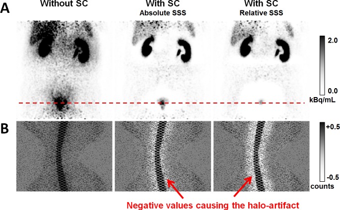
Objectives: Combined positron emission tomography (PET) and magnetic resonance imaging (MRI) targeting the prostate-specific membrane antigen (PSMA) with a 68Ga-labelled PSMA-analog (68Ga-PSMA-11) is discussed as a promising diagnostic method for patients with suspicion or history of prostate cancer. One potential drawback of this method are severe photopenic (halo-) artifacts surrounding the bladder and the kidneys in the scatter-corrected PET images, which have been reported to occur frequently in clinical practice. The goal of this work was to investigate the occurrence and impact of these artifacts and, secondly, to evaluate variants of the standard scatter correction method with regard to halo-artifact suppression.

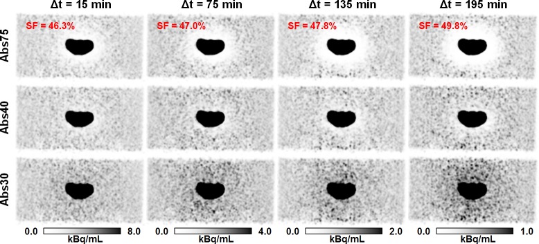
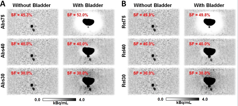
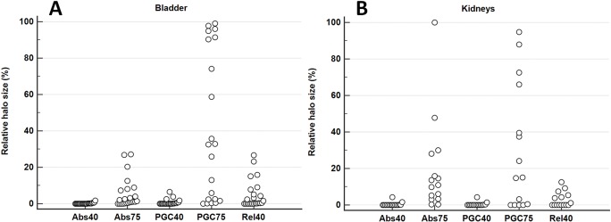
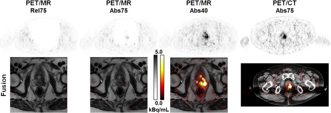
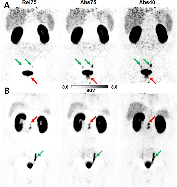
Methods: Experiments using a dedicated pelvis phantom were conducted to investigate whether the halo-artifact is modality-, tracer-, and/or concentration-dependent. Furthermore, 31 patients with history of prostate cancer were selected from an ongoing 68Ga-PSMA-11-PET/MRI study. For each patient, PET raw data were reconstructed employing six different variants of PET scatter correction: absolute scatter scaling, relative scatter scaling, and relative scatter scaling combined with prompt gamma correction, each of which was combined with a maximum scatter fraction (MaxSF) of MaxSF = 75% or MaxSF = 40%. Evaluation of the reconstructed images with regard to halo-artifact suppression was performed both quantitatively using statistical analysis and qualitatively by two independent readers.

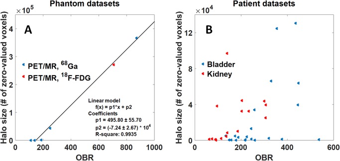
Results: The phantom experiments did not reveal any modality-dependency (PET/MRI vs. PET/CT) or tracer-dependency (68Ga vs. 18F-FDG). Patient- and phantom-based data indicated that halo-artifacts derive from high organ-to-background activity ratios (OBR) between bladder/kidneys and surrounding soft tissue, with a positive correlation between OBR and halo size. Comparing different variants of scatter correction, reducing the maximum scatter fraction from the default value MaxSF = 75% to MaxSF = 40% was found to efficiently suppress halo-artifacts in both phantom and patient data. In 1 of 31 patients, reducing the maximum scatter fraction provided new PET-based information changing the patient's diagnosis.

Conclusion: Halo-artifacts are particularly observed for 68Ga-PSMA-11-PET/MRI due to 1) the biodistribution of the PSMA-11-tracer resulting in large OBRs for bladder and kidneys and 2) inaccurate scatter correction methods currently used in clinical routine, which tend to overestimate the scatter contribution. If not compensated for, 68Ga-PSMA-11 uptake pathologies may be masked by halo-artifacts leading to false-negative diagnoses. Reducing the maximum scatter fraction was found to efficiently suppress halo-artifacts.


Conflict of interest statement

Competing Interests: The authors have declared that no competing interests exist.

Figures

Fig 1
Fig 1. Example for halo-artifacts surrounding the urinary bladder and the kidneys.
(A) PET images in coronal slice orientation for a 59 years old patient with and without scatter correction (SC). Administered activity was 144 MBq of 68Ga-PSMA-11 and the patient was scanned with the mMR 104 min p.i. Absolute SSS resulted in a strongly reduced halo-artifact around the bladder compared to relative SSS. (B) Sinograms corresponding to the direct plane indicated by the dashed red line. Shown are the prompts after subtraction of the estimated scatter. Note the grayscale windowing, which is chosen such that white color indicates negative values.
Fig 2
Fig 2. Comparison of halo-artifact appearance in PET/MR and PET/CT for 68Ga and 18F-FDG.
PET images of the pelvis phantom in coronal slice orientation using absolute SSS with MaxSF = 75%. The administered tracer was either 68Ga or 18F-FDG and scans were performed subsequently in PET/CT (mCT) and PET/MRI (mMR). The mCT data were reconstructed twice: without and with TOF information. A severe halo-artifact surrounding the bladder insert was visible in all six cases.
Fig 3
Fig 3. Effect of reduction of maximum scatter scatter fraction on halo-artifact appearance.
PET images of the pelvis phantom in coronal slice orientation using absolute scatter scaling and both default (MaxSF = 75%) and reduced maximum scatter fraction (MaxSF = 40% or MaxSF = 30%). The administered tracer was 68Ga (33 MBq, background) and 18F-FDG (23 MBq, bladder insert). PET raw data were acquired with the mMR 15, 75, 135, and 195 min p.i., and corresponding OBRs were 108, 136, 184, and 248, respectively. The scatter fraction (SF) increased with increasing time, i.e., increasing OBR A halo-artifact increasing in size over time was observed when the default maximum scatter fraction (MaxSF = 75%) was applied. The halo-artifact was hardly visible for any measurement when using the reduced MaxSF = 40%. Further reduction of MaxSF, e.g., to 30% resulted in activity overestimation surrounding the bladder insert.
Fig 4
Fig 4. Effect of the halo-artifact on lesion quantification.
PET images of the pelvis phantom in coronal slice orientation for the lesion quantification experiment using absolute (A) and relative (B) scatter scaling. Without the bladder insert, both lesions can be clearly identified. With bladder insert, a severe halo-artifact is observed using the default MaxSF = 75%, masking one of the two lesions. Quantitative numbers corresponding to the reconstructions shown are given in Table 1. SF specifies the scatter fraction for each case.
Fig 5
Fig 5. Relation between organ-to-background ratio (OBR) and halo size.
(A) Correlation between OBR and halo size for different phantom experiments using either 68Ga (blue) or 18F-FDG (red) acquired on a PET/MRI system applying the default absolute SSS with MaxSF = 75%. OBR and halo size were fitted against a linear function. (B) Correlation between OBR and halo size for PET/MRI patient data sets for the Abs75 reconstruction. The halo size was calculated for a total number of 21 and 16 patients for bladder (blue) and kidneys (red), respectively. The data show a clear trend to larger halo sizes with increasing OBR.
Fig 6
Fig 6. Evaluation of different scatter correction variants.
For each patient data set, these graphs visualize the relative halo size for the different scatter correction variants compared to Rel75 (100%). A total of 21 patients were evaluated for the bladder (A) and 16 in case of the kidneys (B).
Fig 7
Fig 7. Reducing the maximum scatter fraction may change the patient’s diagnosis.
71 years old patient with biochemical recurrence of a prostate carcinoma (Gleason score 9, determined by biopsy) in the left lobe after primary radiation therapy (74 Gy). Administered activity was 189 MBq 68Ga-PSMA-11 and the patient was scanned with the mMR 179 min p.i. This example demonstrates the clinical impact of the halo-artifact resulting from high uptake ratio in the bladder and very low uptake in the abdominal soft tissue and fat (OBR = 330). In Rel75 and Abs75 reconstructions, the recurrence is not (Rel75) or only faintly (Abs75) detectable, both in the PET (top row) and the fused (bottom row) images. Reducing MaxSF to 40% is mandatory to avoid false-negative diagnosis in the PET component (the MRI component suggested recurrence). Reference is given by PET/CT 1h p.i. (OBR = 109).
Fig 8
Fig 8. Effects of different scatter correction variants on PET quantification.
(A) Patient (52 years, 160 MBq 68Ga-PSMA-11, 188 min p.i.) with prostate carcinoma (red arrow, Gleason score 9, confirmed by surgery). Mean and maximum standardized uptake values SUVmean/SUVmax values evaluated in the pathological lesion indicated by the red arrow: 0/0 (Rel75), 3.0/8.3 (Abs75), 4.3/10.6 (Abs40). (B) Patient (66 years, 237 MBq 68Ga-PSMA-11, 137 min p.i.) with biochemical recurrence after prostatectomy (red arrow, Gleason score 9, confirmed by surgery). SUVmean/SUVmax values evaluated in the pathological lesion indicated by the red arrow: 5.3/8.6 (Rel75), 9.1/14.1 (Abs75), 11.3/17.6 (Abs40). Green arrows indicate physiology (ureters). Standardized 3D-isocontour (40% of SUVmax) was used for SUV quantification.
Fig 9
Fig 9. Effect of arm truncation on halo-artifact appearance.
Attenuation maps and corresponding PET images of a clinical case acquired with a Biograph mMR in coronal slice orientation. In the standard MR-based attenuation maps, arms are truncated. Detruncation of the arms in the attenuation map significantly reduces the size of the halo-artifact for the applied relative (Rel) scaling. Additional prompt gamma correction (PGC) further reduces the halo-size but cannot completely compensate for the artifact, which potentially masks lesions in close vicinity to the bladder. For comparison, the reconstruction obtained with absolute scatter scaling and MaxSF = 40% is shown, which results in a complete halo-artifact suppression.

References

    1. Silver DA, Pellicer I, Fair WR, Heston WWD, Cordon-Cardo C. Prostate-specific membrane antigen expression in normal and malignant human tissues. Clin Cancer Res. 1997;3:81–85. - PubMed
    1. Sweat SD, Pacelli A, Murphy GP, Bostwick DG. Prostate-specific membrane antigen expression is greatest in prostate adenocarcinoma and lymph node metastases. Urology. 1998;52:637–640. - PubMed
    1. Eder M, Eisenhut M, Babich J, Haberkorn U. PSMA as a target for radiolabelled small molecules. Eur J Nucl Med Mol Imaging. 2013;40:819–823. doi: 10.1007/s00259-013-2374-2 - DOI - PMC - PubMed
    1. Eder M, Schäfer M, Bauder-Wüst U, Hull W-E, Wängler C, Mier W, et al. 68Ga-complex lipophilicity and the targeting property of a urea-based PSMA inhibitor for PET imaging. Bioconjug Chem. 2012;23:688–697. doi: 10.1021/bc200279b - DOI - PubMed
    1. Afshar-Oromieh A, Malcher A, Eder M, Eisenhut M, Linhart HG, Hadaschik BA, et al. PET imaging with a [68Ga]gallium-labelled PSMA ligand for the diagnosis of prostate cancer: biodistribution in humans and first evaluation of tumour lesions. Eur J Nucl Med Mol Imaging. 2013;40:486–495. doi: 10.1007/s00259-012-2298-2 - DOI - PubMed