Review
. 2017 Aug 16;95(4):757-778.
doi: 10.1016/j.neuron.2017.06.014.

Toward a Wiring Diagram Understanding of Appetite Control

Affiliations
Review

Toward a Wiring Diagram Understanding of Appetite Control

Mark L Andermann et al. Neuron. .

Abstract

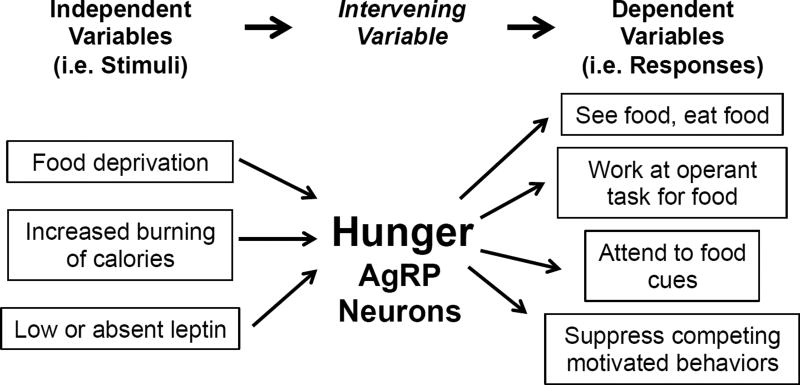
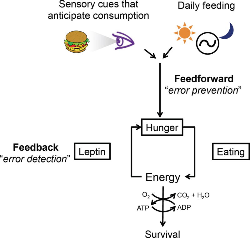
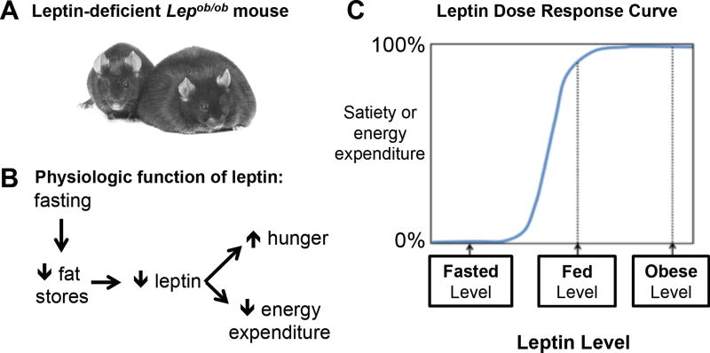
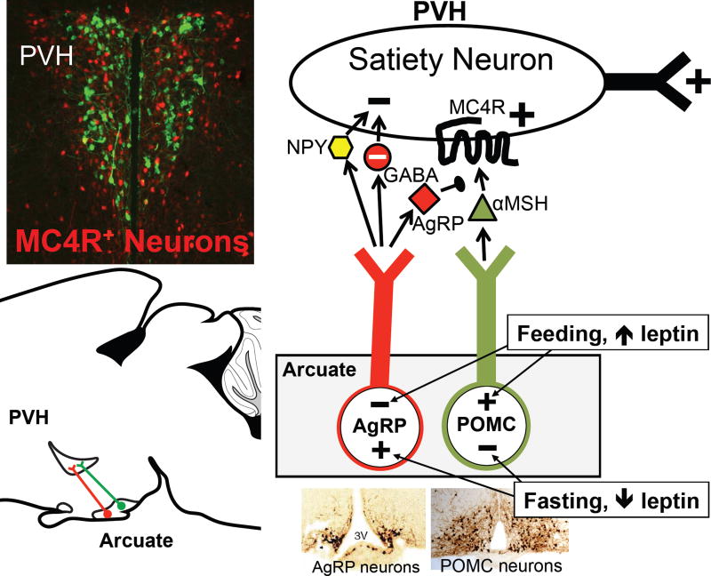
Prior mouse genetic research has set the stage for a deep understanding of appetite regulation. This goal is now being realized through the use of recent technological advances, such as the ability to map connectivity between neurons, manipulate neural activity in real time, and measure neural activity during behavior. Indeed, major progress has been made with regard to meal-related gut control of appetite, arcuate nucleus-based hypothalamic circuits linking energy state to the motivational drive, hunger, and, finally, limbic and cognitive processes that bring about hunger-mediated increases in reward value and perception of food. Unexpected findings are also being made; for example, the rapid regulation of homeostatic neurons by cues that predict future food consumption. The aim of this review is to cover the major underpinnings of appetite regulation, describe recent advances resulting from new technologies, and synthesize these findings into an updated view of appetite regulation.

Keywords: AgRP neurons; POMC neurons; anticipatory regulation; arcuate nucleus; feedforward regulation; food cues; hunger; intervening variable; melanocortin-4 receptor; satiety.


Figures

Figure 1
Figure 1. Hunger and AgRP neurons as the “intervening variable”
The intervening variable concept of motivational drive simplifies the linking of different stimuli (independent variables) with many different responses (dependent variables) (as proposed by Neal Miller (Miller, 1971) and modified by (Berridge, 2004)).
Figure 2
Figure 2. Homeostasis and the role of feedback and feedforward control
Feedback is reactive in that it detects perturbations, i.e. “errors”, and regulates hunger to restore energy balance. Feedforward regulation anticipates future needs and regulates hunger to prevent future disturbances in energy balance.
Figure 3
Figure 3. Satiation and the role of the afferent vagus nerve and NTS
Vagal afferents detect stretch of the gut wall and nutrient-induced release of paracrine signals such as CCK and serotonin (5HT) by gut enteroendocrine cells. These afferents activate neurons in the NTS to bring about satiation.
Figure 4
Figure 4. “Labeled line” transmission by Glp1r- and Gpr65-expressing vagal afferents (Williams et al., 2016)
(A) Vagal afferents marked by expression of Glpr1 detect stretch and, via a vago-vagal reflex, appear to produce stomach contractions. As stomach stretch has been shown to inhibit feeding (Phillips and Powley, 1996), these neurons likely engage NTS neurons that bring about satiation. (B) Vagal afferents marked by expression of Gpr65 detect nutrient-induced release of 5HT, and via a vago-vagal reflex, inhibit stomach contractions. This serves to prevent excessively rapid transit of nutrients from the stomach into the small intestine. The effect of Gpr65-expressing afferents on behavior is presently unknown.
Figure 5
Figure 5. Satiation: NTS → LPBN and beyond
NTS neurons activate LPBNCGRP neurons and this promotes meal termination via projections to the central amygdala (CeA) (Campos et al., 2016; Carter et al., 2013; Roman et al., 2016). NTSCCK neurons also project to the PVH where they promote satiety (D'Agostino et al., 2016). Similarly, NTSGLP1 neurons can promote satiety (Gaykema et al., 2017), perhaps via their projections to the PVH and ARC.
Figure 6
Figure 6. The adipocyte hormone, leptin
Low levels of leptin signal to the brain that fat stores are inadequate (Ahima et al., 1996; Rosenbaum and Leibel, 2014). (A) A control sibling (left) sitting next to a homozygous leptin-deficient, very obese mouse (right). Image courtesy of R.L. Leibel (2008) Int. J. Obes. 32, S98, 2008. (B) Decreased food intake reduces fat stores and lowers blood leptin levels. This is sensed by the brain, which then brings about adaptive changes in appetite and energy expenditure – both aimed at restoring fat stores. (C) A schematized dose response curve for blood leptin levels and their effects on satiety and energy expenditure. The effective range for leptin is between the low levels seen with fasting and the normal levels seen in the ad libitum fed, non-obese state. Levels above this, as occur with obesity or with exogenous leptin treatment, produce little additional effects – i.e. the dose response curve at higher levels of leptin is relatively flat.
Figure 7
Figure 7. The ARC to PVH satiety circuit
See text for details. Upper left insert – PVH neurons. MC4R–expressing neurons (red) were identified by crossing Mc4r-2a–Cre mice with lox-tdTomato reporter mice. The section was counterstained with an antibody against oxytocin (green). Note that oxytocin neurons do not express MC4Rs and that Mc4r–expressing neurons are a minor yet functionally important subset of all PVH neurons.
Figure 8
Figure 8. The complete ARC→ PVH circuit includes ARCVGLUT2 satiety neurons
Slow (AgRP) and fast (NPY and GABA) mediators of hunger are released by one set of neurons, ARCAgRP neruons. Slow (αMSH) and fast (glutamate) mediators of satiety, on the other hand, are released by two parallel-projecting neurons, ARCPOMC and ARCVGLUT2 neurons (Fenselau et al., 2017). αMSH/MC4R signaling in PVHMC4Rneurons causes satiety by two mechanisms: by directly activating the MC4R–PVH neurons and via synaptic plasticity - upregulating excitatory transmission across the ARCVGLUT2 → PVHMC4R synapse (as indicated by the blue line) (Fenselau et al., 2017). PVHMC4R neurons project to the lateral parabrachial nucleus (LPBN) where they promote satiety (Garfield et al., 2015).
Figure 9
Figure 9. Long range afferent regulation of AgRP neurons
(A) Strong excitatory afferents come from the PVH and drive hunger via a reciprocal PVH → ARC — | PVH → satiety circuit (Krashes et al., 2014). Strong inhibition comes from LEPR-expressing neurons in the DMH, and these afferents promote rapid food cue-induced regulation of AgRP neurons (Garfield et al., 2016). The excitatory afferents may also promote rapid, food cue-induced regulation – although this has yet to be tested. (B) AgRP neurons have many dendritic spines and their excitatory synapses are very plastic. Fasting markedly increases spine number and synaptic activity, and these fasting-induced plasticity responses, which require NMDARs on AgRP neurons, contribute to fasting activation of AgRP neurons (Liu et al., 2012). Photo of dendritic spines courtesy of Dong Kong.
Figure 10
Figure 10. Rapid drops in AgRP neuron activity during food presentation
Presentation of a quantity of food sufficient to replenish energy deficit causes a large drop in AgRP neuron activity within seconds (A, black line), albeit not to quiescent levels. If ‘feedforwarD' multisensory signals predicting the consequences of consumption of this food on energy deficit were not available, the drop in firing would only occur over tens of minutes or more (A, blue line), due to systemic feedback (e.g. from increased leptin, etc.). When mice are presented with smaller quantities of food that will only partially restore energy balance, the rapid drop in AgRP activity is proportionately smaller (B). In the limit of very small food rewards (C), AgRP firing should remain elevated and roughly constant across many food presentations.
Figure 11
Figure 11. A more detailed model of feedback and feedforward control
Recent studies implicate AgRP neurons as a key ‘intervening variable’ between estimation of current and future caloric needs and orchestration of diverse food seeking behaviors (compare to Figure 2). Estimates of current energy deficit and upcoming energy balance following consumption are controlled by feedback signals to AgRP neurons (e.g. leptin, ghrelin), as well as feedforward signals (e.g. DMHLepR neuron inputs to AgRP neurons) and direct feedback modulation of these feedforward inputs.
Figure 12
Figure 12. A neural pathway illustrating hunger-dependent processing of food-cue sensory processing
Many studies in humans and rodents implicate the basolateral amygdala (BLA) and insular cortex (InsCtx) in assessing the valence and salience of learned food cues, and addressing the question of expected interoceptive value: how will eating this food make me feel, now and later? If the answer is net positive, then motor circuits are recruited by BLA and InsCtx. This answer is dependent on hunger state (and therefore on tonic input from AgRP neurons, in part via paraventricular thalamus, PVT), and on the salience of learned food cue inputs to BLA (from thalamus and association cortex). The estimation of interoceptive consequences of food consumption in InsCtx is likely shaped by previous experience involving visceral and gustatory input. LP: lateral posterior nucleus of the thalamus. POR: postrhinal cortex. VPL: ventral posterolateral thalamus. VPM: ventral posteromedial thalamus. PBN: parabrachial nucleus. NTS: nucleus of the solitary tract. NAc: nucleus accumbens (Livneh et al., 2017).

References

    1. Ahima RS, Prabakaran D, Mantzoros C, Qu D, Lowell B, Maratos-Flier E, Flier JS. Role of leptin in the neuroendocrine response to fasting. Nature. 1996;382:250–252. - PubMed
    1. Allen LG, Kalra PS, Crowley WR, Kalra SP. Comparison of the effects of neuropeptide Y and adrenergic transmitters on LH release and food intake in male rats. Life Sci. 1985;37:617–623. - PubMed
    1. Aotani D, Ebihara K, Sawamoto N, Kusakabe T, Aizawa-Abe M, Kataoka S, Sakai T, Iogawa H, Ebihara C, Fujikura J, et al. Functional magnetic resonance imaging analysis of food-related brain activity in patients with lipodystrophy undergoing leptin replacement therapy. J Clin Endocrinol Metab. 2012;97:3663–3671. - PMC - PubMed
    1. Aponte Y, Atasoy D, Sternson SM. AGRP neurons are sufficient to orchestrate feeding behavior rapidly and without training. Nat Neurosci. 2011;14:351–355. - PMC - PubMed
    1. Atasoy D, Betley JN, Li WP, Su HH, Sertel SM, Scheffer LK, Simpson JH, Fetter RD, Sternson SM. A genetically specified connectomics approach applied to long-range feeding regulatory circuits. Nat Neurosci. 2014;17:1830–1839. - PMC - PubMed

Substances