. 2017 Aug 22;70(8):958-971.
doi: 10.1016/j.jacc.2017.06.049.

Pharmacological and Activated Fibroblast Targeting of Gβγ-GRK2 After Myocardial Ischemia Attenuates Heart Failure Progression

Affiliations

Pharmacological and Activated Fibroblast Targeting of Gβγ-GRK2 After Myocardial Ischemia Attenuates Heart Failure Progression

Joshua G Travers et al. J Am Coll Cardiol. .

Abstract

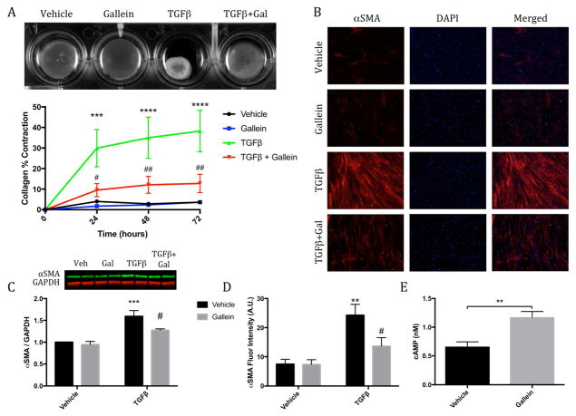
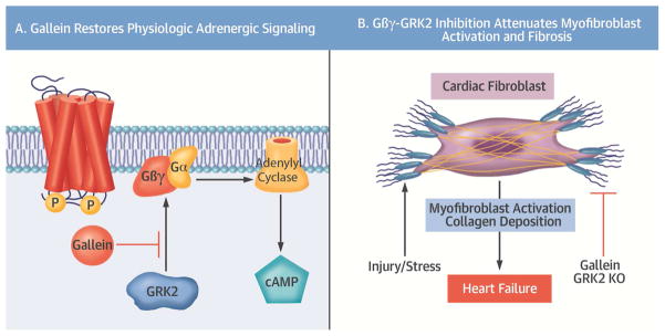
Background: Cardiac fibroblasts are a critical cell population responsible for myocardial extracellular matrix homeostasis. Upon injury or pathological stimulation, these cells transform to an activated myofibroblast state and play a fundamental role in myocardial fibrosis and remodeling. Chronic sympathetic overstimulation, a hallmark of heart failure (HF), induces pathological signaling through G protein βγ (Gβγ) subunits and their interaction with G protein-coupled receptor kinase 2 (GRK2).

Objectives: This study investigated the hypothesis that Gβγ-GRK2 inhibition and/or ablation after myocardial injury would attenuate pathological myofibroblast activation and cardiac remodeling.

Methods: The therapeutic potential of small molecule Gβγ-GRK2 inhibition, alone or in combination with activated fibroblast- or myocyte-specific GRK2 ablation-each initiated after myocardial ischemia-reperfusion (I/R) injury-was investigated to evaluate the possible salutary effects on post-I/R fibroblast activation, pathological remodeling, and cardiac dysfunction.

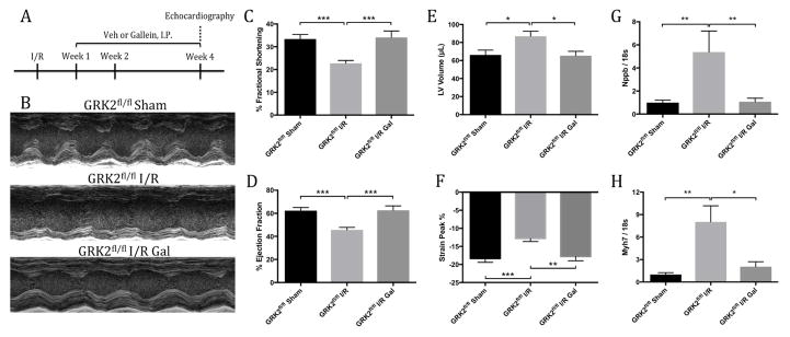
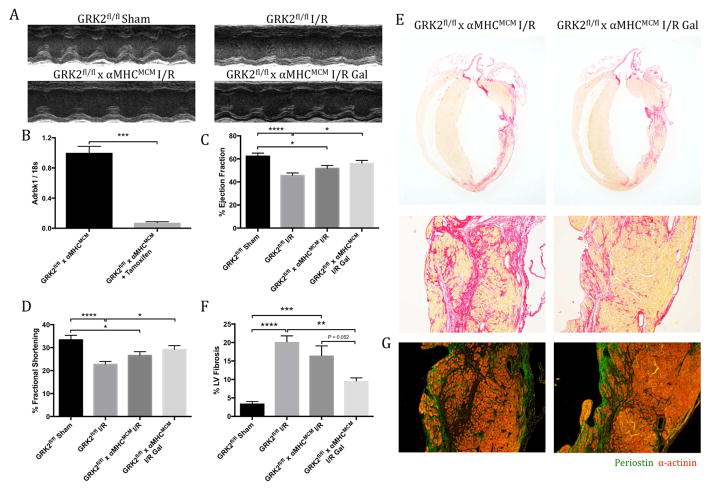
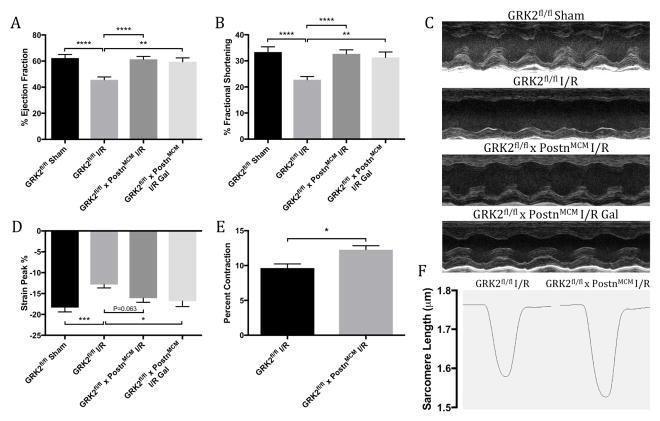
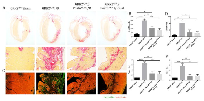
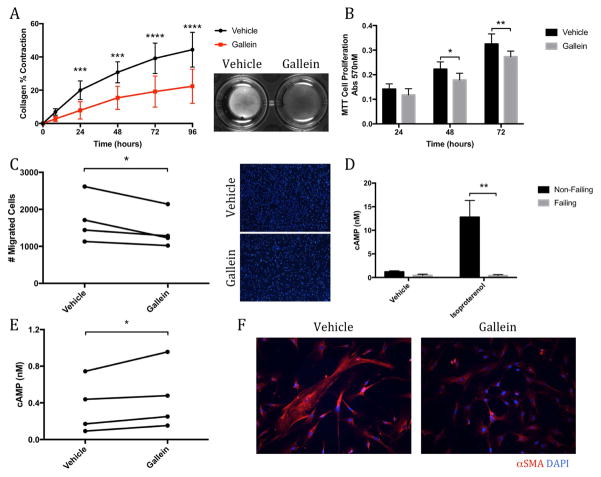
Results: Small molecule Gβγ-GRK2 inhibition initiated 1 week post-injury was cardioprotective in the I/R model of chronic HF, including preservation of cardiac contractility and a reduction in cardiac fibrotic remodeling. Systemic small molecule Gβγ-GRK2 inhibition initiated 1 week post-I/R in cardiomyocyte-restricted GRK2 ablated mice (also post-I/R) still demonstrated significant cardioprotection, which suggested a potential protective role beyond the cardiomyocyte. Inducible ablation of GRK2 in activated fibroblasts (i.e., myofibroblasts) post-I/R injury demonstrated significant functional cardioprotection with reduced myofibroblast transformation and fibrosis. Systemic small molecule Gβγ-GRK2 inhibition initiated 1 week post-I/R provided little to no further protection in mice with ablation of GRK2 in activated fibroblasts alone. Finally, Gβγ-GRK2 inhibition significantly attenuated activation characteristics of failing human cardiac fibroblasts isolated from end-stage HF patients.

Conclusions: These findings suggested consideration of a paradigm shift in the understanding of the therapeutic role of Gβγ-GRK2 inhibition in treating HF and the potential therapeutic role for Gβγ-GRK2 inhibition in limiting pathological myofibroblast activation, interstitial fibrosis, and HF progression.

Keywords: cardiac fibroblast; cardioprotection; fibrosis; remodeling.


Figures

Figure 1
Figure 1. Cardiac Function and HF Marker Expression
(A) Timeline of ischemia-reperfusion (I/R) injury surgery and gallein (Gal) or vehicle (Veh) administration in mice. Effect of G protein-coupled receptor kinase 2 (GRK2) inhibition seen in (B) representative m-mode echocardiographic images and cardiac functional parameters including (C) percent fractional shortening, (D) ejection fraction, (E) left ventricular (LV) end-diastolic volume and (F) peak percentage by strain analysis. Gene expression levels of heart failure (HF) markers (G) Nppb (statistical analysis following log transformation) and (H) Myh7 normalized to 18s ribosomal sub-unit as determined by quantitative polymerase chain reaction (qPCR). *p < 0.05; **p < 0.01; ***p < 0.001.
Figure 2
Figure 2. Fibrotic Scar Formation and Marker Expression
The effect of Gβγ-GRK2 inhibition on fibrotic scar formation (A) was assessed by picrosirius red staining, and (B) quantification was expressed as percentage of collagen deposition over total LV area. (C) Representative images show periostin expression within the ventricular free wall. Messenger ribonucleic acid levels of fibrosis markers (D) Col1a1 and (E) Postn were measured by qPCR and normalized to 18s ribosomal sub-unit. *p < 0.05; **p < 0.01; ***p < 0.001; ****p < 0.0001. Abbreviations as in Figure 1.
Figure 3
Figure 3. Cardiac Function and Fibrosis in Cardiomyocyte-specific GRK2 Knockout Mice Post-I/R
(A) Representative m-mode echocardiographic images showing ventricular contractility. (B) GRK2 transcript expression in cardiomyocytes isolated from GRK2fl/fl x α-MHCMCM mice by qPCR analysis. Function quantified by (C) percent ejection fraction and (D) fractional shortening. Collagen deposition assessed by (E) picrosirius red staining, with the fibrotic area quantified as percentage of collagen deposition over total left ventricular area (F). (G) Representative images of periostin (Postn) expression within the ventricular free wall 4 weeks post-I/R. *p < 0.05; **p < 0.01; ***p < 0.001; ****p < 0.0001. α-MHC = α-myosin heavy chain; other abbreviations as in Figure 1.
Figure 4
Figure 4. Cardiac Function and Cardiomyocyte Contractility in Activated Fibroblast GRK2 Knockout Mice Post-I/R
Cardiac function assessed by echocardiography is shown by (A) percent ejection fraction and (B) fractional shortening 4 weeks post-I/R, with (C) representative long-axis m-mode images at level of papillary muscle. (D) Peak percentage determined by strain analysis. Sarcomeric contractility was assessed in primary cardiomyocytes 4 weeks post-I/R; shown are (E) percent sarcomeric contractility and (F) representative sarcomeric length traces over a single contraction. *p < 0.05; **p < 0.01; ***p < 0.001; ****p < 0.0001. Abbreviations as in Figure 1.
Figure 5
Figure 5. Fibrosis and Fibrotic Marker Gene Expression in Activated Fibroblast GRK2 Knockout Mice Following Injury
Fibrotic scar formation assessed by (A) picrosirius red staining, with (B) quantification expressed as proportion of fibrotic region over total LV area. (C) Representative images of periostin expression within the ventricular free wall. Transcript expression of fibrosis markers (D) Col1a1, (E) Postn, and (F) Fn1, measured by qPCR and normalized to 18s. *p < 0.05; **p < 0.01; ***p < 0.001; ****p < 0.0001. Abbreviations as shown in Figures 1 and 3.
Figure 6
Figure 6. Effect of Gβγ-GRK2 Inhibition on Pathological Myofibroblast Activation
(A) Collagen contractility assay with representative collagen gels showing contraction 72 h after gel release, with the percent collagen gel contraction quantified over a 72-h period. ***p < 0.001 vs. Veh; ****p < 0.0001 vs. Veh; #p < 0.05 vs. transforming growth factor-β (TGF-β); ##p < 0.01 vs. TGF-β. (B) Representative smooth muscle α actin (α-SMA) immunofluorescence images. (C) Western blot analysis of α-SMA expression normalized to glyceraldehyde 3-phosphate dehydrogenase (GAPDH). (D) Quantification of relative α-SMA fluorescence intensity. **p < 0.01 vs. Veh; ***p < 0.001 vs. Veh; #p < 0.05 vs. TGF-β. (E) Production of cyclic adenosine monophosphate (cAMP) measured following 10 min of isoproterenol stimulation. **p < 0.01. Other abbreviations as in Figure 1.
Figure 7
Figure 7. Failing Human Cardiac Fibroblasts: Reduced Activation State
Gallein is shown to reduce the activation state of failing human cardiac fibroblasts. Analysis of (A) collagen gel contraction and (B) cell proliferation by MTT assay in failing human cardiac fibroblasts treated with gallein. *p < 0.05 vs. Veh; **p < 0.01 vs. Veh; ***p < 0.001 vs. Veh; ****p < 0.0001 vs. Veh. (C) Cell migration was measured across a cell-permeable transwell membrane; representative membrane images are shown from which migrated cells were quantified. *p < 0.05. Production of cAMP (D) assessed in normal and failing human cardiac fibroblasts following isoproterenol stimulation. (**p < 0.01 vs. non-failing) and (E) in failing human cardiac fibroblasts treated with or without gallein (*p < 0.05). (F) Representative images of α-SMA immunofluorescence. DAPI = 4',6-diamidino-2-phenylindole; other abbreviations as in Figure 6.
Central Illustration
Central Illustration. Attenuation of Myofibroblast Activation by Gβγ-GRK2 Inhibition
(A) The cardioprotective effects of gallein are mediated through disruption of the interaction between active Gβγsubunits and G protein-coupled receptor kinase 2 (GRK2), thus reducing desensitization and downregulation of β-adrenergic receptors. This restoration of β-adrenergic receptor signaling potentiated production of the second messenger cyclic adenosine monophosphate (cAMP) (A) to reduce fibroblast activation and the resulting fibrotic remodeling, thereby attenuating the progression of heart failure (B). KO = knockout.

Comment in

References

    1. Benjamin EJ, Blaha MJ, Chiuve SE, et al. Heart Disease and Stroke Statistics-2017 Update: A Report From the American Heart Association. Circulation. 2017;135:e146–e603. - PMC - PubMed
    1. Travers JG, Kamal FA, Robbins J, Yutzey KE, Blaxall BC. Cardiac Fibrosis: The Fibroblast Awakens. Circ Res. 2016;118:1021–40. - PMC - PubMed
    1. Sun Y, Weber KT. Animal models of cardiac fibrosis. Methods Mol Med. 2005;117:273–90. - PubMed
    1. Kamal FA, Travers JG, Blaxall BC. G protein-coupled receptor kinases in cardiovascular disease: why “where” matters. Trends Cardiovasc Med. 2012;22:213–9. - PubMed
    1. Li J, Philip JL, Xu X, Theccanat T, Abdur Razzaque M, Akhter SA. beta-Arrestins regulate human cardiac fibroblast transformation and collagen synthesis in adverse ventricular remodeling. J Mol Cell Cardiol. 2014;76:73–83. - PMC - PubMed

MeSH terms