. 2017 Dec;76(12):2095-2103.
doi: 10.1136/annrheumdis-2017-211499. Epub 2017 Aug 17.

B cell OX40L supports T follicular helper cell development and contributes to SLE pathogenesis

Affiliations

B cell OX40L supports T follicular helper cell development and contributes to SLE pathogenesis

Andrea Cortini et al. Ann Rheum Dis. 2017 Dec.

Abstract

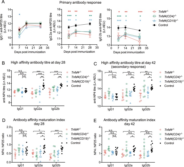
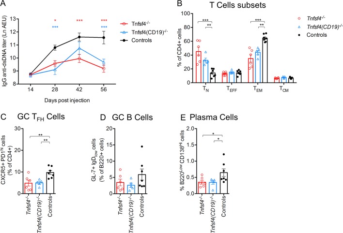
Objectives: TNFSF4 (encodes OX40L) is a susceptibility locus for systemic lupus erythematosus (SLE). Risk alleles increase TNFSF4 expression in cell lines, but the mechanism linking this effect to disease is unclear, and the OX40L-expressing cell types mediating the risk are not clearly established. Blockade of OX40L has been demonstrated to reduce disease severity in several models of autoimmunity, but not in SLE. We sought to investigate its potential therapeutic role in lupus.

Methods: We used a conditional knockout mouse system to investigate the function of OX40L on B and T lymphocytes in systemic autoimmunity.

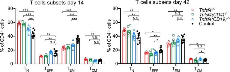
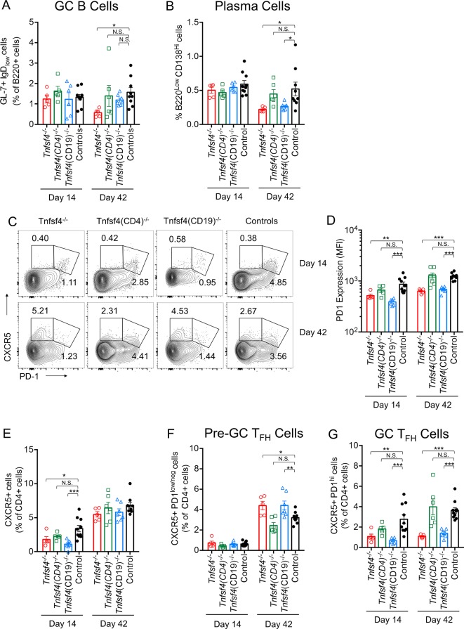
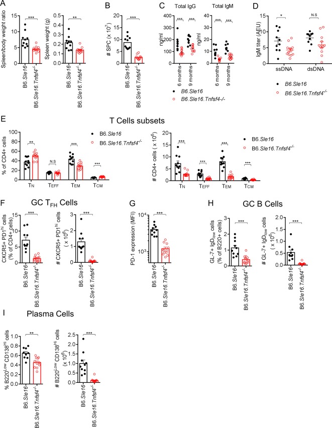
Results: Physiologically, OX40L on both B and T cells contributed to the humoral immune response, but B cell OX40L supported the secondary humoral response and antibody affinity maturation. Our data also indicated that loss of B cell OX40L impeded the generation of splenic T follicular helper cells. We further show that in two models of SLE-a spontaneous congenic model and the H2-IAbm12 graft-versus-host-induced model-loss of B cell OX40L ameliorates the autoimmune phenotype. This improvement was, in each case, accompanied by a decline in T follicular helper cell numbers. Importantly, the germline knockout did not exhibit a markedly different phenotype from the B cell knockout in these models.

Conclusions: These findings contribute to a model in which genetically determined increased OX40L expression promotes human SLE by several mechanisms, contingent on its cellular expression. The improvement in pathology in two models of systemic autoimmunity indicates that OX40L is an excellent therapeutic target in SLE.

Keywords: B cells; OX40L; T follicular helper cells; autoantibodies; systemic lupus erythematosus.


Conflict of interest statement

Competing interests: None declared.

Figures

Figure 1
Figure 1
OX40L in T cell-dependent humoral response. Wild-type controls, Tnfsf4−/−, Tnfsf4fl/fl(CD19)−/− and Tnfsf4fl/fl(CD4)−/− mice were immunised with NP-CGG in CFA and reimmunised on day 35 with NP-CGG in IFA. Sera were collected on days 7, 14 and 28 for the primary response and on day 42 for the secondary response. (A) Titres of NP-specific low-affinity antibody measured with NP25-BSA. (B) Titres of NP-specific high-affinity antibodies on day 28 measured with NP4-BSA. (C) Titres of NP-specific high-affinity antibodies on day 42 measured with NP4-BSA. (D) Affinity maturation index calculated as ratio of the titres of IgG detected with NP4-BSA to those with NP25-BSA on day 28. (E) Affinity maturation index calculated as the ratio of the titres of IgG detected with NP4-BSA to those with NP25-BSA on day 42. Each symbol represents an individual mouse; dots in (A) and bars in (B–E) indicate the mean titre, each being shown mean±SEM. AEU is arbitrary ELISA unit; N.S. is not significant; *p<0.05, **p<0.01 and ***p<0.001 (A, two-way ANOVA; B–E, one-way ANOVA). ANOVA, analysis of variance; CFA, complete Freund’s adjuvant; IFA, incomplete Freund’s adjuvant; NP-CGG, nitrophenylacetyl-chicken gamma globulin; NP25-BSA, NP4-BSA, 4-Hydroxy-3-nitrophenylacetyl hapten conjugated to bovine serum albumin.
Figure 2
Figure 2
Role of OX40L in T cell activation. Wild-type controls, Tnfsf4−/−, Tnfsf4(CD19)−/− and Tnfsf4(CD4)−/− mice were immunised with NP-CGG in CFA and reimmunised on day 35 with NP-CGG in IFA. Spleens were taken and analysed by FACS either on day 14 or day 42. Quantification of naïve (CD4+, CD62L+, CD44low) T cells, effector (CD4+, CD62Llow, CD44low) T cells, effector/memory (CD4+, CD62Llow/neg, CD44hi) T cells and central/memory (CD4+, CD62L+, CD44hi) T cells on day 14 (left) and day 42 (right). Each symbol represents an individual mouse. Bars indicate the mean±SEM. N.S., not significant; *p<0.05, **p<0.01 and ***p<0.001 (one-way analysis of variance). CFA, complete Freund’s adjuvant; FACS, fluorescence-activated cell sorting; IFA, incomplete Freund’s adjuvant; NP-CGG, nitrophenylacetyl-chicken gamma globulin.
Figure 3
Figure 3
OX40L function in GC reaction. Wild-type controls, Tnfsf4−/−, Tnfsf4(CD19)−/− and Tnfsf4(CD4)−/− mice were immunised with NP-CGG in CFA and reimmunised on day 35 with NP-CGG in IFA. Spleens were taken and analysed by FACS either on day 14 or day 42. (A) Frequency of GC B cell (B220+, GL7+, IgD−) presented as frequency among the B220+ population. (B) Percentage of plasma cells (B220low, CD138hi). (C) Gating of T follicular helper (TFH) (CD4+, CXCR5+, PD-1hi) and pre-T follicular helper (pre-TFH) (CD4+, CXCR5+, PD-1Low/neg) cells. (D) PD-1 expression level in CD4+ cells assessed by FACS. (E) Frequency of CXCR5+ cells presented as frequency among the CD4+ population. (F) Quantification of pre-GC TFH and (G) GC-TFH cells as gated in (C) presented as frequency among the CD4+ population. Each symbol represents an individual mouse. Bars indicate the mean±SEM, N.S., not significant; *p<0.05, **p<0.01 and ***p<0.001 (one-way analysis of variance). CFA, complete Freund’s adjuvant; FACS, fluorescence-activated cell sorting; GC, germinal centre; IFA, incomplete Freund’s adjuvant; NP-CGG, nitrophenylacetyl-chicken gamma globulin.
Figure 4
Figure 4
OX40L deficiency ameliorates the phenotype of B6.Sle16 lupus-prone mice. Comparison between female B6.Sle16 and B6.Sle16.Tnfsf4−/− female mice at 9 months of age. (A) Quantitation of spleen/body weight ratio and spleen weight. (B) Absolute number of cells per spleen. (C) Serum level of IgG and IgM at 6 and 9 months. (D) Titre of IgM anti-dsDNA and anti-ssDNA. (E) Quantitation of naïve (TN) (CD4+, CD62L+, CD44low), (TEFF) effector (CD4+, CD62Llow, CD44low), TEM effector/memory (CD4+, CD62Llow/neg, CD44hi) and TCM central/memory (CD4+, CD62L+, CD44hi) T cells expressed as a percentage of CD4+ cells and absolute number. (F) GC TFH cells (CD4+, CXCR5+, PD-1hi) presented as frequency among the CD4+ population and absolute number. (G) PD-1 expression level in CD4+ cells assessed by FACS. (H) GC B cell (B220+, GL7+, IgD−) presented as frequency among the B220+ population and absolute number. (I) Percentage and absolute number of plasma cells (B220low, CD138hi). Each symbol represents an individual mouse. Bars indicate the mean±SEM N.S., not significant; *p<0.05, **p<0.01 and ***p<0.001 (t-test). dsDNA, double-stranded deoxyribonucleic acid; FACS, fluorescence-activated cell sorting; GC, germinal centre; ssDNA, single-stranded deoxyribonucleic acid.
Figure 5
Figure 5
OX40L deficiency diminishes anti-dsDNA antibody production in the cGvHD model. Female wild-type controls, Tnfsf4−/− and Tnfsf4(CD19)−/− mice were injected intraperitoneally with 5×10⁷ splenocytes from B6.H2bm12 female mice. Sera were collected on days 14, 28, 42 and 56. On day 56, spleens were collected and analysed by FACS. (A) Titre of IgG anti-dsDNA in the sera of injected mice at different time points. (B) Quantification of naïve (TN) (CD4+, CD62L+, CD44low), (TEFF) effector (CD4+, CD62Llow, CD44low), (TEM) effector/memory (CD4+, CD62Llow/neg, CD44hi) and (TCM) central/memory (CD4+, CD62L+, CD44hi) T cells expressed as percentage of CD4+ cells. (C) Quantification of GC TFH cells (CD4+, CXCR5+, PD-1hi) presented as frequency among the CD4+ population. (D) Frequency of GC B cells (B220+,GL7+, IgD−) presented as frequency among the B220+ population. (E) Percentage of plasma cells (B220low, CD138hi). Each symbol represents data from an individual mouse. Bars indicate the mean±SEM. N.S., not significant; *p<0.05, **p<0.01 and ***p<0.001 (one-way analysis of variance). cGvHD, chronic graft-versus-host-disease; dsDNA, double-stranded deoxyribonucleic acid; FACS, fluorescence-activated cell sorting; GC, germinal centre.

Comment in

References

    1. Mohan C, Putterman C. Genetics and pathogenesis of systemic lupus erythematosus and lupus nephritis. Nat Rev Nephrol 2015;11:329–41. 10.1038/nrneph.2015.33 - DOI - PubMed
    1. Tsokos GC. Systemic lupus erythematosus. N Engl J Med 2011;365:2110–21. 10.1056/NEJMra1100359 - DOI - PubMed
    1. Harley JB, Alarcón-Riquelme ME, Criswell LA, et al. . Genome-wide association scan in women with systemic lupus erythematosus identifies susceptibility variants in ITGAM, PXK, KIAA1542 and other loci. Nat Genet 2008;40:204–10. 10.1038/ng.81 - DOI - PMC - PubMed
    1. Bentham J, Morris DL, Graham DSC, et al. . Genetic association analyses implicate aberrant regulation of innate and adaptive immunity genes in the pathogenesis of systemic lupus erythematosus. Nat Genet 2015;47:1457–64. 10.1038/ng.3434 - DOI - PMC - PubMed
    1. Cunninghame Graham DS, Graham RR, Manku H, et al. . Polymorphism at the TNF superfamily gene TNFSF4 confers susceptibility to systemic lupus erythematosus. Nat Genet 2008;40:83–9. 10.1038/ng.2007.47 - DOI - PMC - PubMed