. 2017 Aug 18;357(6352):703-706.
doi: 10.1126/science.aan2556.

Vinculin forms a directionally asymmetric catch bond with F-actin

Affiliations

Vinculin forms a directionally asymmetric catch bond with F-actin

Derek L Huang et al. Science. .

Abstract

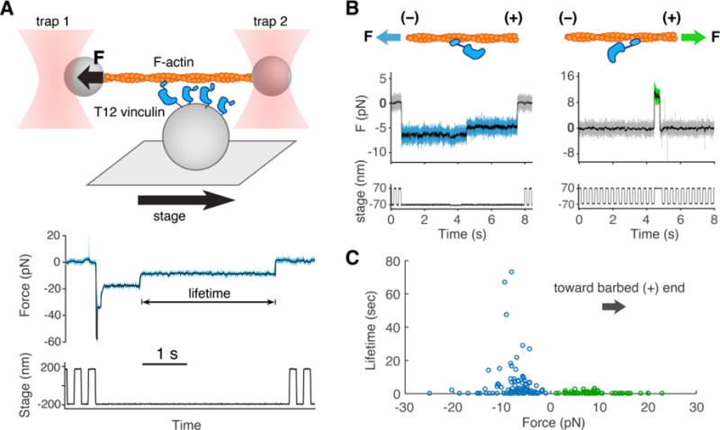
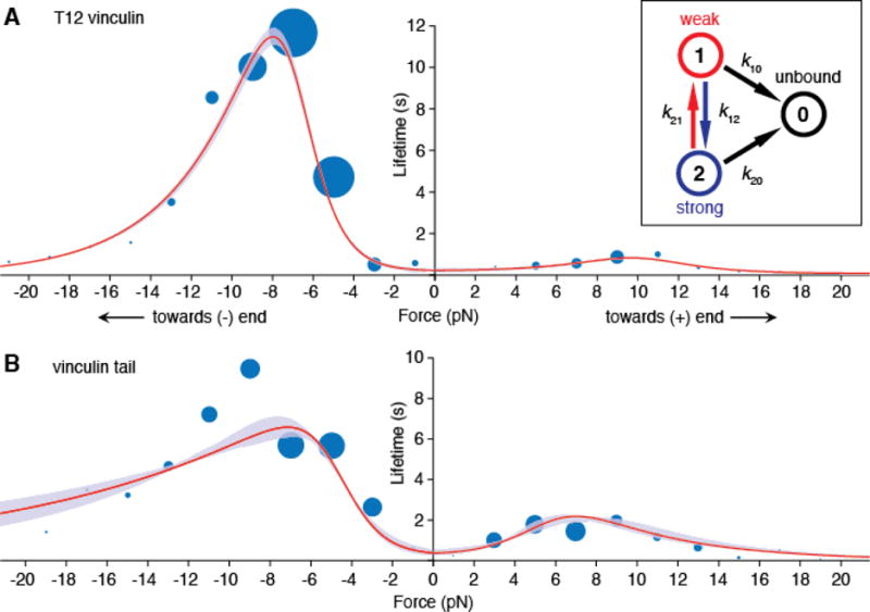
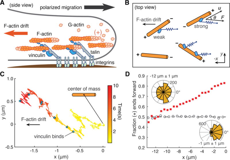
Vinculin is an actin-binding protein thought to reinforce cell-cell and cell-matrix adhesions. However, how mechanical load affects the vinculin-F-actin bond is unclear. Using a single-molecule optical trap assay, we found that vinculin forms a force-dependent catch bond with F-actin through its tail domain, but with lifetimes that depend strongly on the direction of the applied force. Force toward the pointed (-) end of the actin filament resulted in a bond that was maximally stable at 8 piconewtons, with a mean lifetime (12 seconds) 10 times as long as the mean lifetime when force was applied toward the barbed (+) end. A computational model of lamellipodial actin dynamics suggests that the directionality of the vinculin-F-actin bond could establish long-range order in the actin cytoskeleton. The directional and force-stabilized binding of vinculin to F-actin may be a mechanism by which adhesion complexes maintain front-rear asymmetry in migrating cells.


Figures

Fig. 1
Fig. 1. The optical trap (OT) assay measures vinculin/F-actin bond lifetimes under load
(A) (top) An actin filament attached to two microspheres is held taut by two OTs near a platform bead with vinculin on its surface. A motorized stage moves the platform back and forth. Vinculin binding results in the displacement of one of the optically trapped microspheres. (bottom) Both OTs exert force on the actin filament. Here, we plot the summed force, i.e. the total force transmitted from both traps to the vinculin molecule(s) (22) vs. time, decimated from 40 kHz to 4 kHz (light blue) and median filtered with a 100 point moving window (overlaid in black). If force surpasses a defined threshold, stage motion halts until detachment of the bound vinculin molecule(s). (B) Representative traces where force on T12 vinculin is directed toward the pointed (-) end (blue), or the barbed (+) end (green) of the actin filament. (C) Binding events in which the force on T12 vinculin was directed towards the filament’s pointed (−) end (blue circles, n = 102, mean lifetime of 5.6 s) or barbed (+) end (green circles, n = 65, mean lifetime of 0.54 s).
Fig. 2
Fig. 2. A two bound-state directional catch bond model of T12 vinculin and vinculin tail (Vt) binding lifetimes
(A) Mean actin binding lifetimes and model of best fit for T12 vinculin (n = 728) and (B) Vt (n = 702). A subset of these events are collected from filaments of known polarity, and the rest are assigned a polarity using a 2D KS test (22). In each 2 pN bin, the area of each blue circle is proportional to the number of events. Red curves show mean lifetimes predicted by the two bound-state catch bond model (inset), and purple envelopes indicate 95% confidence intervals (CIs) for the fit, obtained by parametric bootstrapping. The model was constrained to possess a mean lifetime at zero force that was less than or equal to the lifetime measured using the low-force binding assay (Fig. S1). Survival plots of the data and models are shown in Fig. S8 for T12 vinculin and Fig. S9 for Vt.
Fig. 3
Fig. 3. Computational model of actin dynamics in a cell protrusion
(A) Long-range order of the actin cytoskeleton (orange) induced by adhesion proteins, including vinculin, may reinforce polarized cell migration. (B) Computational model: actin filaments (orange) undergo 2D Brownian diffusion with a drift velocity. The effect of vinculin on filament motion is modeled as a simple spring that dampens diffusion and resists the drift velocity (22). Directionality in the model depends on the angle θ between the filament’s long axis (u) and the force vector (F). (C) Sample trace of the 2D position of a filament’s center of mass over time. Blue dots indicate times at which an additional vinculin binds. (D) Filaments start at x = 0 μm with random initial angle. Red squares show the fraction of filaments with barbed (+) end facing forward after 60 s. Grey circles show a control simulation in which vinculin’s actin-binding kinetics do not depend on actin polarity. Filaments oriented with barbed (+) end to the right are defined as forward facing (angles −90° to 90°). Inset, top left: angles of filaments within x = −12 μm +/− 1 μm. Inset, bottom right: angles of filaments within x = −1 μm +/− 1 μm. For each condition, n = 224000 filaments.

Comment in

References

    1. Gumbiner BM. Cell adhesion: the molecular basis of tissue architecture and morphogenesis. Cell. 1996;84:345–357. - PubMed
    1. Pruitt BL, Dunn AR, Weis WI, Nelson WJ. Mechano-Transduction: From Molecules to Tissues. PLoS Biol. 2014;12:e1001996. - PMC - PubMed
    1. Montell DJ. Morphogenetic cell movements: diversity from modular mechanical properties. Science. 2008;322:1502–1505. - PubMed
    1. Mutsaers SE, Bishop JE, McGrouther G, Laurent GJ. Mechanisms of tissue repair: from wound healing to fibrosis. Int J Biochem Cell Biol. 1997;29:5–17. - PubMed
    1. Friedl P, Wolf K. Tumour-cell invasion and migration: diversity and escape mechanisms. Nat Rev Cancer. 2003;3:362–374. - PubMed

Publication types