

Elimination of the male reproductive tract in the female embryo is promoted by COUP-TFII in mice
- PMID: 28818950
- PMCID: PMC5713893
- DOI: 10.1126/science.aai9136
Elimination of the male reproductive tract in the female embryo is promoted by COUP-TFII in mice
Abstract
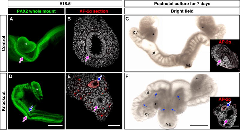
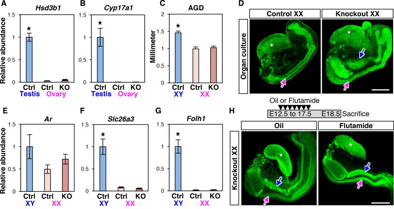
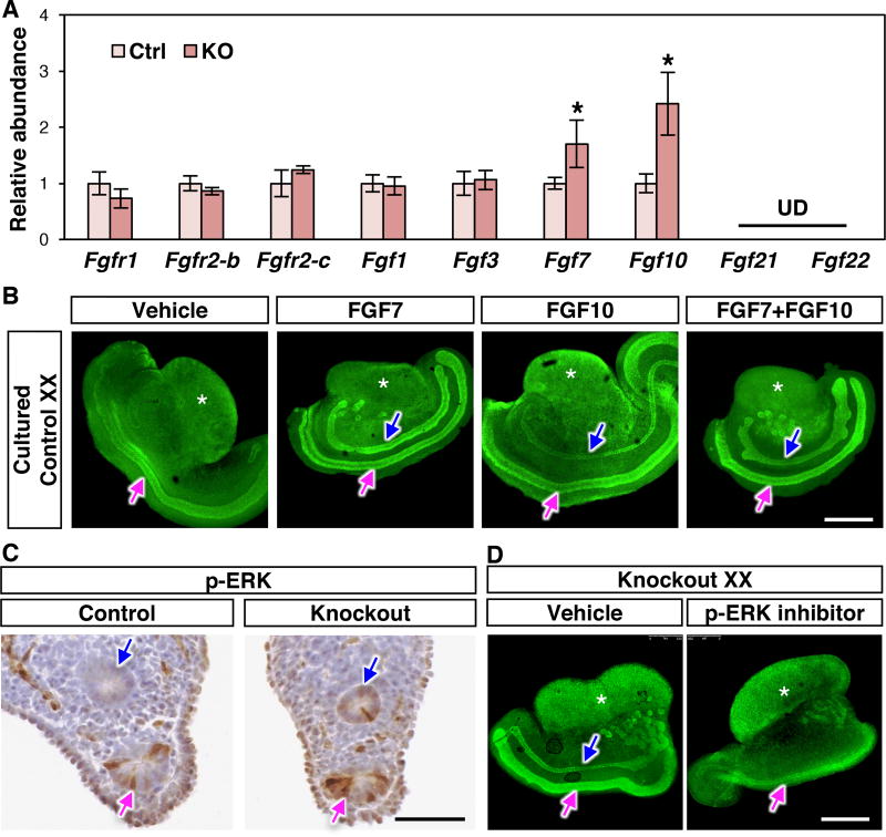
The sexual differentiation paradigm contends that the female pattern of the reproductive system is established by default because the male reproductive tracts (Wolffian ducts) in the female degenerate owing to a lack of androgen. Here, we discovered that female mouse embryos lacking Coup-tfII (chicken ovalbumin upstream promoter transcription factor II) in the Wolffian duct mesenchyme became intersex-possessing both female and male reproductive tracts. Retention of Wolffian ducts was not caused by ectopic androgen production or action. Instead, enhanced phosphorylated extracellular signal-regulated kinase signaling in Wolffian duct epithelium was responsible for the retention of male structures in an androgen-independent manner. We thus suggest that elimination of Wolffian ducts in female embryos is actively promoted by COUP-TFII, which suppresses a mesenchyme-epithelium cross-talk responsible for Wolffian duct maintenance.
Copyright © 2017 The Authors, some rights reserved; exclusive licensee American Association for the Advancement of Science. No claim to original U.S. Government Works.
Figures
Comment in
-
Ductal sex determination.Science. 2017 Aug 18;357(6352):648. doi: 10.1126/science.aao2630. Science. 2017. PMID: 28818931 No abstract available.
-
Regression of the male reproductive tract in the female embryo is regulated by the orphan nuclear receptor COUP-TFII.Biol Reprod. 2017 Oct 1;97(4):517-518. doi: 10.1093/biolre/iox109. Biol Reprod. 2017. PMID: 29025035 No abstract available.
References
-
- Kobayashi A, Behringer RR. Developmental genetics of the female reproductive tract in mammals. Nat Rev Genet. 2003;4:969–980. - PubMed
-
- Jost A. Recherches sur la différenciation sexuelle de l’embryon de lapin. III. Rôle des gonades foetales dans la différenciation sexuelle somatique. Arch Anat Microsc Morph Exp. 1947;36:271–315.
-
- Jost A. Problems of Fetal Endocrinology - the Gonadal and Hypophyseal Hormones. Recent Prog Horm Res. 1953;8:379–418. - PubMed
-
- Cunha GR, et al. Normal and abnormal development of the male urogenital tract. Role of androgens, mesenchymal-epithelial interactions, and growth factors. J Androl. 1992;13:465–475. - PubMed
Publication types
MeSH terms
Substances
Grants and funding
LinkOut - more resources
Full Text Sources
Other Literature Sources
Molecular Biology Databases
