. 2017 Sep 29;357(6358):1416-1420.
doi: 10.1126/science.aao0535. Epub 2017 Aug 17.

Mutations in the promoter of the telomerase gene TERT contribute to tumorigenesis by a two-step mechanism

Affiliations

Mutations in the promoter of the telomerase gene TERT contribute to tumorigenesis by a two-step mechanism

Kunitoshi Chiba et al. Science. .

Abstract

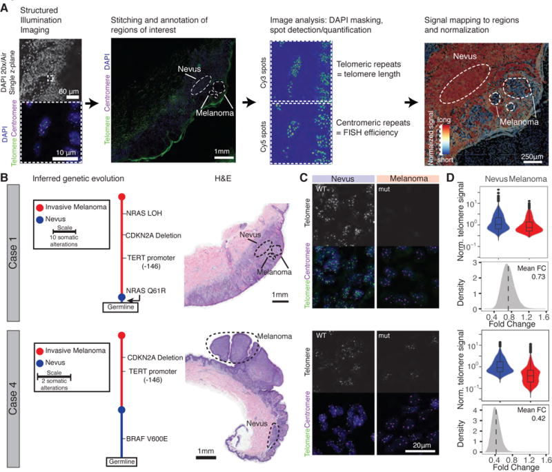
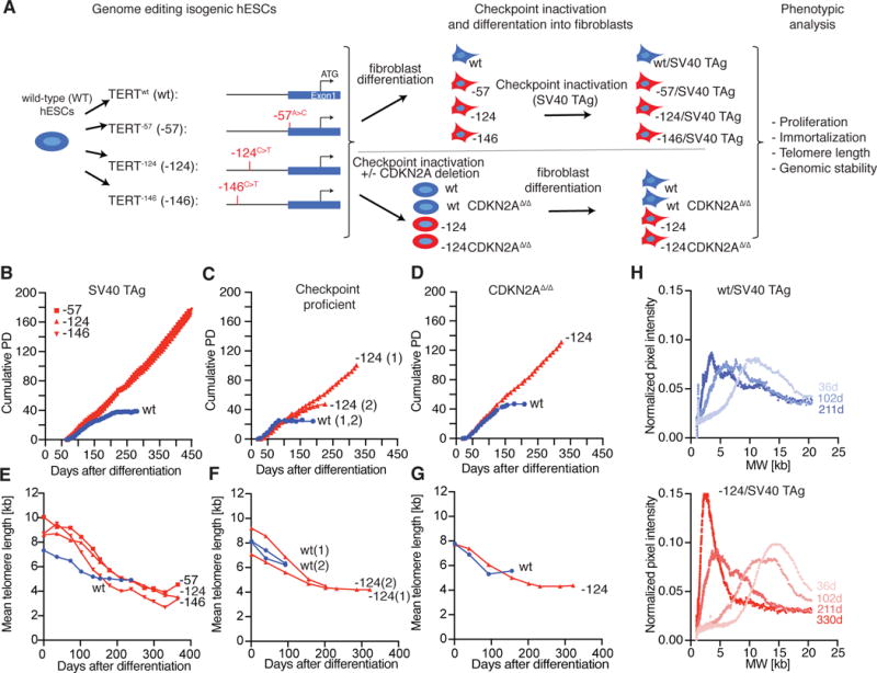
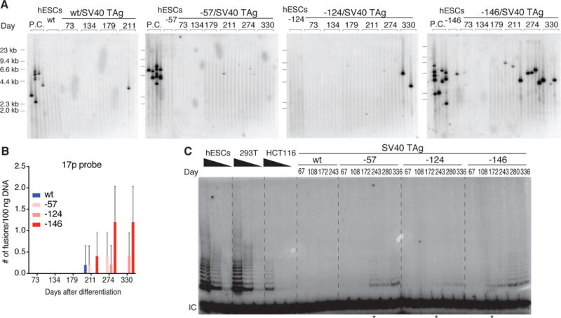
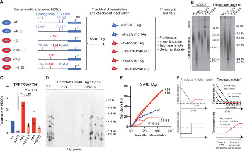
TERT promoter mutations (TPMs) are the most common noncoding mutations in cancer. The timing and consequences of TPMs have not been fully established. Here, we show that TPMs acquired at the transition from benign nevus to malignant melanoma do not support telomere maintenance. In vitro experiments revealed that TPMs do not prevent telomere attrition, resulting in cells with critically short and unprotected telomeres. Immortalization by TPMs requires a gradual up-regulation of telomerase, coinciding with telomere fusions. These data suggest that TPMs contribute to tumorigenesis by promoting immortalization and genomic instability in two phases. In an initial phase, TPMs do not prevent bulk telomere shortening but extend cellular life span by healing the shortest telomeres. In the second phase, the critically short telomeres lead to genome instability and telomerase is further up-regulated to sustain cell proliferation.


Figures

Fig. 1
Fig. 1. Telomeres shorten during progression from nevus to melanoma despite acquisition of a TPM
(A) Schematic overview of workflow. Melanoma progression samples from four patients were imaged using structured illumination microscopy. Acquired images were stitched and regions of interest were annotated. Telomeric and centromeric repeat FISH signals were detected and mapped back on pre-defined regions of interest. The relative telomere length was calculated by normalization to the closest centromeric signals. (B) The genetic evolution of each progression case, rooted at the germline shows how each case evolves from a wild-type nevus to a melanoma with TPM (nevus in blue, melanoma in red). Adjacent is the H&E staining of the analyzed sections of each case (Scale bar 1mm). (C) Magnification of the FISH image resolving individual telomere and centromere spots in nevus/melanoma area (DAPI = blue, telomere = green, centromere = magenta). TPM status is noted as wild-type (WT) or mutant (mut) (Scale bar 20 μm). (D) Violin plots of normalized telomere signal (telomere/centromere) with an inset boxplot indicating the median and quartiles of the signal. Histograms show the fold change of telomere spot intensity after random sampling (fold change melanoma/nevus).
Fig. 2
Fig. 2. TPMs support cellular immortalization in vitro but do not prevent telomere shortening
(A) Experimental overview: Isogenic hESCs with the TPMs were differentiated into fibroblasts. To inactivate cell cycle and DNA damage checkpoints, either CDKN2A function was deleted in hESCs prior differentiation or fibroblasts were infected with SV40 TAg. (B-D) Growth curves of cumulative PDs over days after differentiation. (B) SV40 TAg fibroblasts with (red, -57, -124, -146) or without (blue, wt) TPMs (C) DNA damage checkpoint proficient cells with (red, -124) or without (blue, wt) TPM. (1), (2) indicate two independent experiments. (D) CDKN2AΔ/Δ cells with (red, -124) or without (blue, wt) TPM (E-G) Quantification of mean telomere length over time after differentiation. (H) Accumulation of shorter telomeres over time shown by visualization of telomere length distribution of images shown in Fig. S3A. Quantification of the normalized pixel intensity over molecular weight per lane for the indicated time points after differentiation.
Fig. 3
Fig. 3. TPMs do not fully protect against genomic instability and mutant cells gradually increase telomerase expression
(A) Detection of interchromosomal telomere fusions over time in SV40 TAg fibroblasts by PCR of specific subtelomeric regions. Fusions were detected by probes against the 17p subtelomeric regions (23). DNA from cells in crisis served as a positive control (P.C.: TERT, p53 and p16 triple knockout fibroblasts cultured into crisis). (B) Quantification of fusion events shown in Fig. 3A. (C) Telomerase activity assay of the indicated cell lines over time. IC: internal control. * indicates time points when telomerase activity became detectable.
Fig. 4
Fig. 4. Short telomeres and low telomerase levels protect cells from immortalization by TPMs
(A) Schematic overview of allelic series of cells carrying TPMs (red) and/or endogenous ETS site mutations (E3: mutated TC>GA, blue) in the TERT promoter. E3 refers to the simultaneous mutations of all three endogenous ETS sites (-191, -97 and -92 from the translational start site: ATG). (B) Telomere length analysis for hESCs and fibroblasts with TPMs and ETS mutation combinations. (C) TERT expression normalized to GAPDH relative to wt hESC. Error bars: SEM, two-sided student t-test, n = 3. (D) Telomere fusions detected in -124 and -124 E3 fibroblasts with 17p. (E) Growth curve of fibroblasts with or without TPMs and/or ETS mutations. (F) Telomere length (top) and TERT expression (bottom) in a revised model for immortalization by TPMs. In the classic crisis model, TERT expression is acquired during crisis as a single event (left panel). In the biphasic model (right panel), TPMs can be acquired in a telomere length independent manner at any time (Phase 1). In phase 2 cells are required to gradually upregulate telomerase. Due to unprotected telomeres cells can acquire genomic instability.

Comment in

References

    1. Greider CW, Blackburn EH. Identification of a specific telomere terminal transferase activity in Tetrahymena extracts. Cell. 1985;43:405–413. - PubMed
    1. Greider CW, Blackburn EH. The telomere terminal transferase of Tetrahymena is a ribonucleoprotein enzyme with two kinds of primer specificity. Cell. 1987;51:887–898. - PubMed
    1. Horn S, et al. TERT promoter mutations in familial and sporadic melanoma. Science. 2013;339:959–961. - PubMed
    1. Huang FW, et al. Highly recurrent TERT promoter mutations in human melanoma. Science. 2013;339:957–959. - PMC - PubMed
    1. Jafri MA, Ansari SA, Alqahtani MH, Shay JW. Roles of telomeres and telomerase in cancer, and advances in telomerase-targeted therapies. Genome Med. 2016;8:69. - PMC - PubMed

Publication types