Review
. 2017 Aug 17;474(17):2953-2976.
doi: 10.1042/BCJ20160783.

The AAA+ ATPase p97, a cellular multitool

Affiliations
Review

The AAA+ ATPase p97, a cellular multitool

Lasse Stach et al. Biochem J. .

Abstract

The AAA+ (ATPases associated with diverse cellular activities) ATPase p97 is essential to a wide range of cellular functions, including endoplasmic reticulum-associated degradation, membrane fusion, NF-κB (nuclear factor kappa-light-chain-enhancer of activated B cells) activation and chromatin-associated processes, which are regulated by ubiquitination. p97 acts downstream from ubiquitin signaling events and utilizes the energy from ATP hydrolysis to extract its substrate proteins from cellular structures or multiprotein complexes. A multitude of p97 cofactors have evolved which are essential to p97 function. Ubiquitin-interacting domains and p97-binding domains combine to form bi-functional cofactors, whose complexes with p97 enable the enzyme to interact with a wide range of ubiquitinated substrates. A set of mutations in p97 have been shown to cause the multisystem proteinopathy inclusion body myopathy associated with Paget's disease of bone and frontotemporal dementia. In addition, p97 inhibition has been identified as a promising approach to provoke proteotoxic stress in tumors. In this review, we will describe the cellular processes governed by p97, how the cofactors interact with both p97 and its ubiquitinated substrates, p97 enzymology and the current status in developing p97 inhibitors for cancer therapy.


Conflict of interest statement

The Authors declare that there are no competing interests associated with the manuscript.

Figures
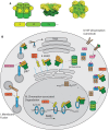
Figure 1.
Figure 1.. Cellular processes governed by p97.
(A) Top: schematic of the p97 architecture, side and top views of the cryo-EM structure of ADP-bound p97 (PDB: 5FTK). Bottom: domain organization of p97, color-coded as above. (B) Well-characterized cellular process requiring p97 activity. (I) Post-mitotic membrane fusion via the p97/p47 pathway. (II) Endoplasmic reticulum-associated degradation. (III) Removal of KU70/80 from chromatin. (IV) NF-κB activation via the canonical pathway. Proteins are color-coded according to their function. p97 and the proteasome are in green, ubiquitinated p97 substrates are blue, cognate E3 ubiquitin ligases decorating p97 substrates are red, phosphorylation signaling proteins are in pink, ubiquitin is in yellow and other proteins are in brown.
Figure 2.
Figure 2.. Table of ubiquitin-associated p97 cofactors.
Left: domain organization of cofactors, color-coded according to domain function, organized according to p97-interacting motif. Middle: cellular processes the cofactors have been implicated in. Right: types of polyubiquitin the cofactors have been associated with or possesses activity against.
Figure 3.
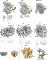
Figure 3.. Structural details of p97-cofactor interactions.
(A) Top: crystal structures of p97 N-domains (gray) in complex with cofactor (gold). FAF1 UBX (PDB: 3QQ8), OTU1 UBX-L (PDB: 4KDL), Npl4 UBX-L (PDB: 2PJH), gp78 VIM (PDB: 3TIW), Rhbdl4 VBM (PDB: 5EPP) and UFD1 SHP (PDB: 5C1B). Conserved residues are indicated. Below: sequence alignment of the conserved interacting motifs, first line in the alignment shows the sequence of the protein shown in the structure above. (B) Composite model of UBX (red), UBX-L (yellow), VIM (green), VBM (blue) and SHP (pink) overlayed on the cryo-EM structure of ADP-bound ATP (5FTK). (C) Crystal structure of PNGase PUB domain (gold) in complex with p97 C-terminal peptide (gray) (PDB: 2HPJ). (D) Overlay of the VIM and VBM complex structures, with the VIM and VBMs rainbow colored from N-terminus (blue) to C-terminus (red). N-domains in gray.
Figure 4.
Figure 4.. Enzymology of p97.
(A) EM structures of p97. ADP bound (PDB: 5FTK), ATPγS (PDB: 5FTN), in complex with FAF1 (EMDB-2319) and the IBMPFD mutant A232E (EMDB-2038). (B) Left: conservation of D1 and D2 pore loop residues in p97, Cdc48 and VAT. Right: structure of pore loops with residues highlighted on table on the left shown as sticks (PDB: 5FTN). (C) Models of p97 conformations in different nucleotide states. p97 domains color-coded as before. IBMPFD mutant positions highlighted in purple. (D) Model for p97 activity. p97 forms a complex with UFD1–NPL4 and a polyubiquitinated substrate. Additional cofactors containing DUB activity remove polyubiquitin leaving K48-chains only. The substrate is then threaded through the central pore of p97 and unfolded.
Figure 5.
Figure 5.. p97 inhibitors.
(A) Table of a selection of known p97 inhibitors, including name, chemical structure, IC50 against p97 ATPase activity and ability to cause an antiproliferative effect in a cell-based assay, e.g. CHOP activation, EC50 of cell death against cancer cell lines A549 and HCT116. (B) Cryo-EM structure of p97 in complex with the UPCDC30245 inhibitor (pink) (PDB: 5FTJ).

References

    1. Erzberger J.P. and Berger J.M. (2006) Evolutionary relationships and structural mechanisms of AAA+ proteins. Annu. Rev. Biophys. Biomol. Struct. 35, 93–114 doi:10.1146/annurev.biophys.35.040405.101933 - DOI - PubMed
    1. DeLaBarre B. and Brunger A.T. (2003) Complete structure of p97/valosin-containing protein reveals communication between nucleotide domains. Nat. Struct. Biol. 10, 856–863 doi:10.1038/nsb972 - DOI - PubMed
    1. Huyton T., Pye V.E., Briggs L.C., Flynn T.C., Beuron F., Kondo H. et al. (2003) The crystal structure of murine p97/VCP at 3.6 Å. J. Struct. Biol. 144, 337–348 doi:10.1016/j.jsb.2003.10.007 - DOI - PubMed
    1. Barthelme D. and Sauer R.T. (2016) Origin and functional evolution of the Cdc48/p97/VCP AAA+ protein unfolding and remodeling machine. J. Mol. Biol. 428(9 Pt B), 1861–1869 doi:10.1016/j.jmb.2015.11.015 - DOI - PMC - PubMed
    1. Peters J.M., Walsh M.J. and Franke W.W. (1990) An abundant and ubiquitous homo-oligomeric ring-shaped ATPase particle related to the putative vesicle fusion proteins Sec18p and NSF. EMBO J. 9, 1757–1767 PMID: - PMC - PubMed

Publication types

MeSH terms