. 2017 Sep;5(9):767-777.
doi: 10.1158/2326-6066.CIR-16-0365. Epub 2017 Aug 17.

The Tumor Microenvironment Regulates Sensitivity of Murine Lung Tumors to PD-1/PD-L1 Antibody Blockade

Affiliations

The Tumor Microenvironment Regulates Sensitivity of Murine Lung Tumors to PD-1/PD-L1 Antibody Blockade

Howard Y Li et al. Cancer Immunol Res. 2017 Sep.

Abstract

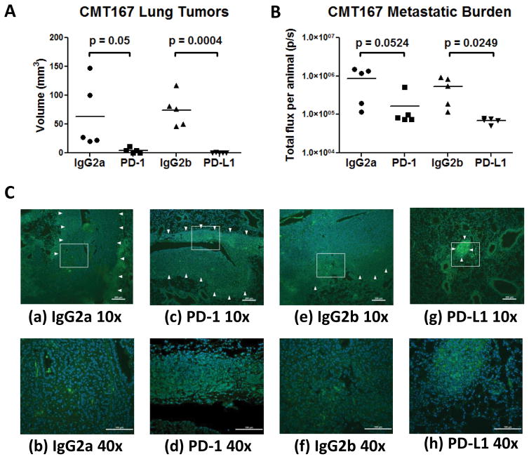
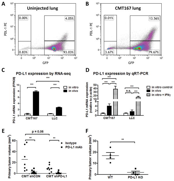
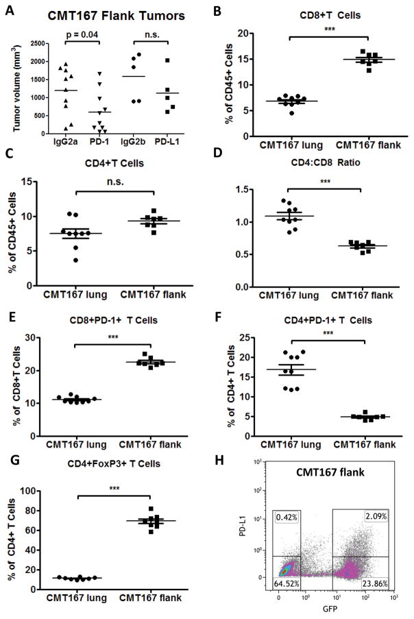
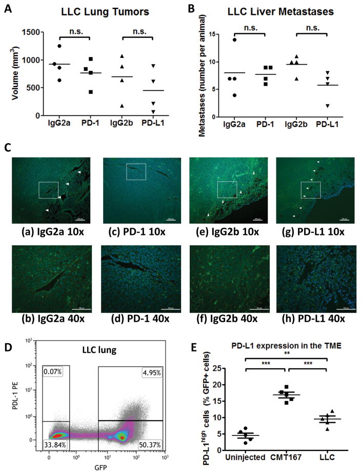
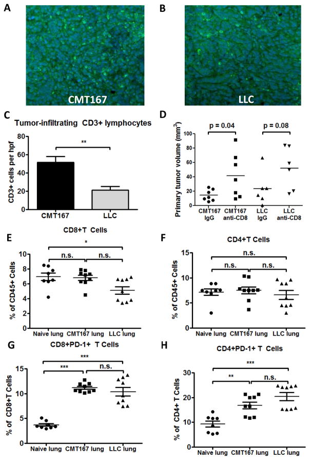
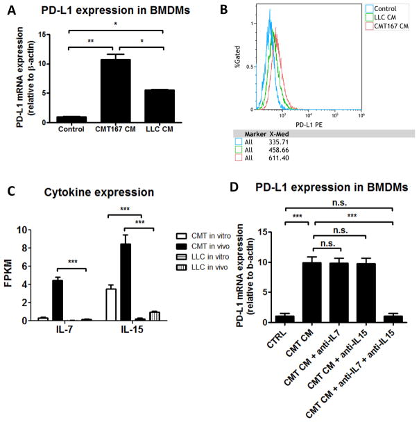
Immune checkpoint inhibitors targeting the interaction between programmed cell death-1 (PD-1) and its ligand PD-L1 induce tumor regression in a subset of non-small cell lung cancer patients. However, clinical response rates are less than 25%. Evaluation of combinations of immunotherapy with existing therapies requires appropriate preclinical animal models. In this study, murine lung cancer cells (CMT167 and LLC) were implanted either orthotopically in the lung or subcutaneously in syngeneic mice, and response to anti-PD-1/PD-L1 therapy was determined. Anti-PD-1/PD-L1 therapy inhibited CMT167 orthotopic lung tumors by 95%. The same treatments inhibited CMT167 subcutaneous tumors by only 30% and LLC orthotopic lung tumors by 35%. CMT167 subcutaneous tumors had more Foxp3+ CD4+ T cells and fewer PD-1+ CD4+ T cells compared with CMT167 orthotopic tumors. Flow cytometric analysis also demonstrated increased abundance of PD-L1high cells in the tumor microenvironment in CMT167 tumor-bearing lungs compared with CMT167 subcutaneous tumors or LLC tumor-bearing lungs. Silencing PD-L1 expression in CMT167 cells resulted in smaller orthotopic tumors that remained sensitive to anti-PD-L1 therapy, whereas implantation of CMT167 cells into PD-L1- mice blocked orthotopic tumor growth, indicating a role for PD-L1 in both the cancer cell and the microenvironment. These findings indicate that the response of cancer cells to immunotherapy will be determined by both intrinsic properties of the cancer cells and specific interactions with the microenvironment. Experimental models that accurately recapitulate the lung tumor microenvironment are useful for evaluation of immunotherapeutic agents. Cancer Immunol Res; 5(9); 767-77. ©2017 AACR.


Figures

Fig. 1
Fig. 1
PD-1 and PD-L1 antibody blockade inhibit growth of CMT167 orthotopic lung tumors. CMT167 orthotopic tumor-bearing mice were injected with anti-PD-1, anti-PD-L1, or isotype control antibody (n = 5 for each group). Effects of anti-PD-1/anti-PD-L1 therapy on (A) CMT167 primary tumor volume and (B) metastatic burden in the other lung lobes. Each point represents an individual mouse. (C) Representative CD3 immunostaining (FITC, green) of sections from mice treated with control IgG2a (a and b), anti-PD-1 (c and d), control IgG2b (e and f), and anti-PD-L1 (g and h). Nuclei were stained with DAPI (blue); tumor borders are indicated by arrowheads. (a,c,e,g) Low power image (10x), scale bar = 200 μm; (b,d,f,h) high power detail of boxed regions from low power images (40x), scale bar = 100 μm.
Fig. 2
Fig. 2
PD-L1 expression is upregulated on both cancer cells and in the TME in CMT167 orthotopic lung tumors. Representative flow cytometric plot from uninjected mice (A) and CMT167 tumor-bearing mice (B). Similar results were obtained in 5 independent experiments, with 2 mice in each pool. (C) PD-L1 mRNA expression from cancer cells recovered from tumor-bearing mice was analyzed by RNA-seq and compared to cancer cells in culture. Each RNA isolation represents a pool of 4–5 mice. (D) PD-L1 mRNA expression from cancer cells recovered from tumor-bearing mice was determined by qRT-PCR and compared with cells cultured in vitro, either in the presence or absence of IFNγ (3 separate RNA isolations). (E) Effects of depleting PD-L1 in CMT167 cells on primary tumor size. CMT shCON isotype n = 7, CMT shCON PD-L1 mAb n = 8, CMT shPD-L1 isotype n = 10, CMT shPD-L1 PD-L1 mAb n = 10. (F) CMT167 cells were implanted in the left lungs of wild-type C57BL/6 mice and PD-L1 KO mice (n = 4 for each group). Statistically significant differences are indicated as determined by one-way ANOVA (C–E) or Student unpaired t test (F), ** = P < 0.01, *** = P < 0.001.
Fig. 3
Fig. 3
CMT167 subcutaneous tumors induce a T-cell response that is skewed toward CD8+ T cells and are less sensitive than CMT167 orthotopic tumors to anti-PD-1/PD-L1 therapy. (A) CMT167 subcutaneous tumor-bearing mice were injected with anti-PD-1, anti-PD-L1, or isotype control antibody (n = 10 for IgG2a and anti-PD-1 groups, n = 5 for IgG2b and anti-PD-L1 groups). CMT167 orthotopic tumor-bearing lungs (n = 9) and CMT167 subcutaneous tumors (n = 7) were analyzed by flow cytometry for (B) CD8+ T cells, (C) CD4+ T cells, (D) CD4:CD8 ratio, (E) PD-1 expression on CD8+ T cells, (F) PD-1 expression on CD4+ T cells, and (G) Foxp3 expression in CD4+ T cells. Statistically significant differences are indicated as determined by Student unpaired t test; ** = P < 0.01, *** = P < 0.001. (H) PD-L1 expression was analyzed by flow cytometry in CMT167 subcutaneous tumors. Similar results were obtained in five independent mice.
Fig. 4
Fig. 4
LLC orthotopic tumors are less sensitive than CMT167 orthotopic tumors to anti-PD-1/PD-L1 therapy. LLC orthotopic tumor-bearing mice were injected with anti-PD-1, anti-PD-L1, or isotype control antibody (n = 4 for each group). Effects of anti-PD-1/anti-PD-L1 therapy on (A) LLC primary tumor volume and (B) number of liver metastases. (C) Representative CD3 immunostaining of tissue sections from mice treated with control IgG2a (a and b), anti-PD-1 (c and d), control IgG2b (e and f), and anti-PD-L1 (g and h). Nuclei were stained with DAPI (blue); tumor borders are indicated by arrowheads. (a, c, e, g) Low power image (10x), scale bar = 200 μm; (b, d, f, h) high power detail of boxed regions from low power images (40x), scale bar = 100 μm. (D) Representative flow cytometric plot from LLC tumor-bearing mice. Similar results were obtained in 5 independent experiments consisting of 2 mice each. (E) The percentage of GFP+PD-L1high cells in mice implanted with CMT167 tumors or LLC tumors was compared to uninjected mice. Each point represents an individual animal (n = 5 for each group). ** = P < 0.01, *** = P < 0.001.
Fig. 5
Fig. 5
Both CMT167 orthotopic lung tumors and LLC orthotopic lung tumors generate an adaptive immune response and induce PD-1 expression on T cells. Tissue sections from CMT167 (representative image shown in Fig. 5A) and LLC (representative image shown in Fig. 5B) tumor-bearing mice were stained for CD3 (FITC, green) and nuclei were stained with DAPI (blue). Magnification x20. (C) Quantification of tumor-infiltrating lymphocytes in CMT167 tumors (n = 9) and LLC tumors (n = 6). (D) Effects of CD8+ T cell immunodepletion on growth of CMT167 (n = 7 each group) and LLC orthotopic tumors (n = 6 each group). CMT167 (n = 9, same mice as in Fig. 3B–F) or LLC tumor-bearing lungs (n = 9) were analyzed by flow cytometry and compared with lungs from naive mice (n = 8) for (E) CD8+ T cells, (F) CD4+ T cells, (G) PD-1 expression on CD8+ T cells, and (H) PD-1 expression on CD4+ T cells. Statistically significant differences are indicated as determined by Student unpaired t test (C-D) or by one-way ANOVA (E-H); * = P < 0.05, ** = P < 0.01, *** = P < 0.001.
Fig. 6
Fig. 6
Conditioned media (CM) from CMT167 cells induces higher levels of PD-L1 in bone marrow derived macrophages than CM from LLC cells. (A) Bone marrow derived macrophages were treated with CM from CMT167 cells or LLC cells. Data represent the mean of 3 experiments using 3 separate isolations of CM. (B) A representative overlay of PD-L1 expression is shown from untreated BMDMs (blue), BMDMs treated with LLC CM (green), and BMDMs treated with CMT167 CM (red). Similar results were obtained in 3 independent experiments using 3 separate isolations of CM. (C) IL7 and IL15 mRNA expression was determined by RNA-seq from CMT167 cells and LLC cells, both in vitro and in vivo. (D) Effects of IL7 and IL15 neutralization on upregulation of PD-L1 on BMDMs induced by CMT167 CM. Data represent the mean and SEM from 3 independent experiments. Statistically significant differences are indicated as determined by one-way ANOVA; * = P < 0.05, ** = P < 0.01, *** = P < 0.001.

References

    1. Jin HT, Ahmed R, Okazaki T. Role of PD-1 in regulating T-cell immunity. Current topics in microbiology and immunology. 2011;350:17–37. - PubMed
    1. Pardoll DM. The blockade of immune checkpoints in cancer immunotherapy. Nature reviews Cancer. 2012;12(4):252–64. - PMC - PubMed
    1. Borghaei H, Paz-Ares L, Horn L, Spigel DR, Steins M, Ready NE, et al. Nivolumab versus Docetaxel in Advanced Nonsquamous Non-Small-Cell Lung Cancer. The New England journal of medicine. 2015 - PMC - PubMed
    1. Brahmer J, Reckamp KL, Baas P, Crino L, Eberhardt WE, Poddubskaya E, et al. Nivolumab versus Docetaxel in Advanced Squamous-Cell Non-Small-Cell Lung Cancer. The New England journal of medicine. 2015;373(2):123–35. - PMC - PubMed
    1. Gettinger SN, Horn L, Gandhi L, Spigel DR, Antonia SJ, Rizvi NA, et al. Overall Survival and Long-Term Safety of Nivolumab (Anti-Programmed Death 1 Antibody, BMS-936558, ONO-4538) in Patients With Previously Treated Advanced Non-Small-Cell Lung Cancer. Journal of clinical oncology : official journal of the American Society of Clinical Oncology. 2015;33(18):2004–12. - PMC - PubMed

Publication types

MeSH terms