

Der f 31, a novel allergen from Dermatophagoides farinae, activates epithelial cells and enhances lung-resident group 2 innate lymphoid cells
- PMID: 28819104
- PMCID: PMC5561047
- DOI: 10.1038/s41598-017-04878-0
Der f 31, a novel allergen from Dermatophagoides farinae, activates epithelial cells and enhances lung-resident group 2 innate lymphoid cells
Abstract
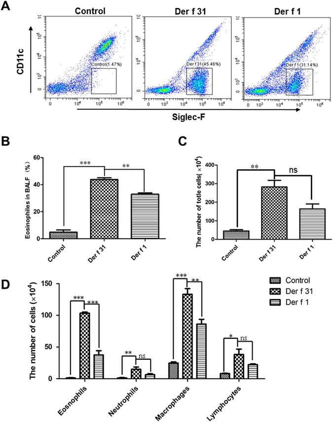
Airway epithelial cell-derived thymic stromal lymphopoietin (TSLP) and IL-33 can enhance lung-resident group 2 innate lymphoid cells (ILC2s), and they play an important role in the development of allergic diseases. This study tests the hypothesis that Der f 31 (Dermatophagoides farinae-31), an allergen, modulates airway epithelial cell functions and increases the frequency of lung ILC2s. Our previous research identified cofilin (Der f 31) as a novel allergen. In this study, we found that recombinant Der f 31 (r-Der f 31) upregulated the expression of co-stimulatory molecules in DCs and promoted Th2-skewed polarization. The levels of TSLP and IL-33 in epithelial cells were upregulated by r-Der f 31 via the activation of Toll-like receptor 2. Furthermore, in in vivo studies, r-Der f 31 induced eosinophil-like airway allergy and increased the number of lung-resident ILC2s. In summary, Der f 31 can modulate the functions of airway epithelial cells and increase levels of lung-resident ILC2s.
Conflict of interest statement
The authors declare that they have no competing interests.
Figures


Similar articles
-
Identification of α-tubulin, Der f 33, as a novel allergen from Dermatophagoides farinae.Immunobiology. 2016 Aug;221(8):911-7. doi: 10.1016/j.imbio.2016.03.004. Epub 2016 Mar 18. Immunobiology. 2016. PMID: 27067709
-
Identification of a novel cofilin-related molecule (Der f 31) as an allergen from Dermatophagoides farinae.Immunobiology. 2018 Feb;223(2):246-251. doi: 10.1016/j.imbio.2017.10.013. Epub 2017 Oct 5. Immunobiology. 2018. PMID: 29102047
-
Der f 38 Is a Novel TLR4-Binding Allergen Related to Allergy Pathogenesis from Dermatophagoides farinae.Int J Mol Sci. 2021 Aug 5;22(16):8440. doi: 10.3390/ijms22168440. Int J Mol Sci. 2021. PMID: 34445142 Free PMC article.
-
Benzo(a)pyrene facilitates dermatophagoides group 1 (Der f 1)-induced epithelial cytokine release through aryl hydrocarbon receptor in asthma.Allergy. 2019 Sep;74(9):1675-1690. doi: 10.1111/all.13784. Epub 2019 Jun 19. Allergy. 2019. PMID: 30982974 Free PMC article.
-
House dust mite Dermatophagoides farinae augments proinflammatory mediator productions and accessory function of alveolar macrophages: implications for allergic sensitization and inflammation.J Immunol. 2003 Jan 1;170(1):528-36. doi: 10.4049/jimmunol.170.1.528. J Immunol. 2003. PMID: 12496440
Cited by
-
Characterization of Innate Immune Responses to House Dust Mite Allergens: Pitfalls and Limitations.Front Allergy. 2021 Mar 11;2:662378. doi: 10.3389/falgy.2021.662378. eCollection 2021. Front Allergy. 2021. PMID: 35386970 Free PMC article. Review.
-
CD157 Confers Host Resistance to Mycobacterium tuberculosis via TLR2-CD157-PKCzeta-Induced Reactive Oxygen Species Production.mBio. 2019 Aug 27;10(4):e01949-19. doi: 10.1128/mBio.01949-19. mBio. 2019. PMID: 31455656 Free PMC article.
-
Bcl11b Regulates IL-17 Through the TGF-β/Smad Pathway in HDM-Induced Asthma.Allergy Asthma Immunol Res. 2018 Sep;10(5):543-554. doi: 10.4168/aair.2018.10.5.543. Allergy Asthma Immunol Res. 2018. PMID: 30088373 Free PMC article.
-
Exosomes Derived from Dermatophagoides farinae Induce Allergic Airway Inflammation.Microbiol Spectr. 2023 Aug 17;11(4):e0505422. doi: 10.1128/spectrum.05054-22. Epub 2023 Jun 14. Microbiol Spectr. 2023. PMID: 37314339 Free PMC article.
-
Increased expression of type 2 innate lymphoid cells in pediatric patients with allergic rhinitis.Exp Ther Med. 2020 Jan;19(1):735-740. doi: 10.3892/etm.2019.8235. Epub 2019 Nov 22. Exp Ther Med. 2020. PMID: 31853326 Free PMC article.
References
Publication types
MeSH terms
Substances
LinkOut - more resources
Full Text Sources
Other Literature Sources
