. 2017 Aug 18;8(1):290.
doi: 10.1038/s41467-017-00359-0.

Landscape of submitochondrial protein distribution

Affiliations

Landscape of submitochondrial protein distribution

F-Nora Vögtle et al. Nat Commun. .

Abstract

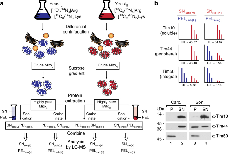
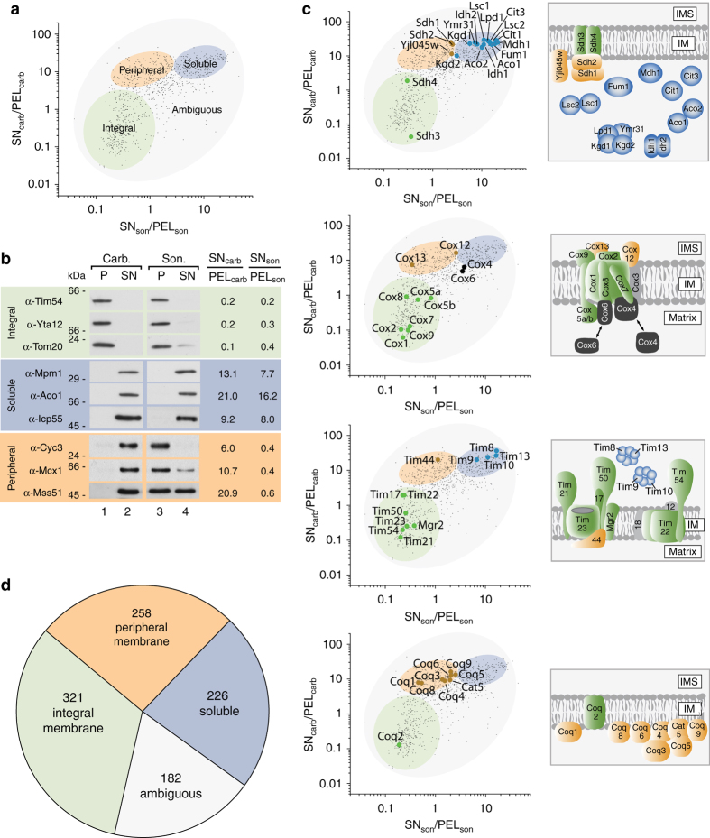
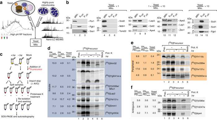
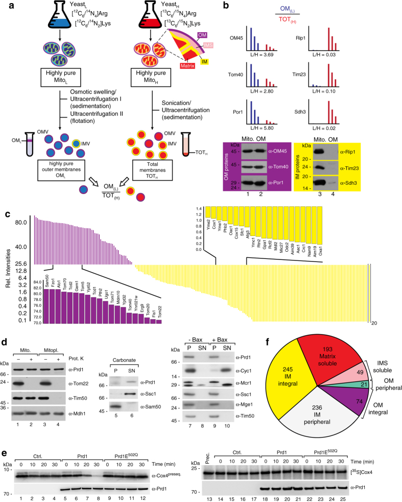
The mitochondrial proteome comprises ~1000 (yeast)-1500 (human) different proteins, which are distributed into four different subcompartments. The sublocalization of these proteins within the organelle in most cases remains poorly defined. Here we describe an integrated approach combining stable isotope labeling, various protein enrichment and extraction strategies and quantitative mass spectrometry to produce a quantitative map of submitochondrial protein distribution in S. cerevisiae. This quantitative landscape enables a proteome-wide classification of 986 proteins into soluble, peripheral, and integral mitochondrial membrane proteins, and the assignment of 818 proteins into the four subcompartments: outer membrane, inner membrane, intermembrane space, or matrix. We also identified 206 proteins that were not previously annotated as localized to mitochondria. Furthermore, the protease Prd1, misannotated as intermembrane space protein, could be re-assigned and characterized as a presequence peptide degrading enzyme in the matrix.Protein localization plays an important role in the regulation of cellular physiology. Here the authors use an integrated proteomics approach to localize proteins to the mitochondria and provide a detailed map of their specific localization within the organelle.


Conflict of interest statement

The authors declare no competing financial interests.

Figures

Fig. 1
Fig. 1
Experimental design for global profiling of peripheral membrane, integral membrane, and soluble proteins in highly pure yeast mitochondria. a Highly pure mitochondria were isolated from yeast cultures grown in the presence of either “light” (L) or “heavy” (H) amino acids (Arg, Lys). Protein extraction was performed based on sonication to separate membrane proteins (PELson) from soluble proteins (SNson) or carbonate treatment, which results in a pellet containing integral membrane proteins (PELcarb) and a supernatant with peripheral membrane and soluble proteins (SNcarb). Respective SN/PEL SILAC ratios were determined by LC-MS. carb, carbonate; son, sonication. b Exemplary SILAC results a for Tim proteins representing the three different protein classes analyzed, confirmed by immunoblotting
Fig. 2
Fig. 2
Mapping of mitochondrial proteins into different classes. a Separation of the mitochondrial proteome into an integral membrane, a peripheral membrane, a soluble, and an ambiguous fraction. b Validation of map positions for several mitochondrial proteins by immunoblotting. c Map data correlation with known submitochondrial localization of components of the translocase of the inner membrane (TIM), the citrate cycle machinery, complex IV of the respiratory chain, and the Coenzyme Q biosynthesis apparatus, , –. Proteins in gray could not be mapped by LC-MS. d Distribution of the mitochondrial proteome into the indicated classes
Fig. 3
Fig. 3
Validation of mitochondrial localization for novel proteins. a Quantitative assessment of protein abundance in total yeast cells and purified mitochondria based on spectral counting (normalized abundance factor). Resulting Yeast/Mito ratios indicate presence of proteins either exclusively in mitochondria or in multiple cellular compartments. b Immunoblot analysis of cellular fractions containing pure mitochondria (Mito), enriched cytosolic proteins (S100), or microsomal proteins (P100). Por1, mitochondrial marker; Pgk1, cytosolic marker; Sss1, ER marker. long, long exposure time; short, short exposure time. c Schematic overview of in organello import reactions to validate mitochondrial localization. [35S]labeled precursors of candidate proteins were generated by in vitro transcription/translation and incubated with isolated mitochondria in the presence or absence of the membrane potential (Δψ) or without mitochondria (Mock). Import reaction was terminated by depletion of the membrane potential and samples were treated with Proteinase K where indicated to remove non-imported precursors. Samples were analyzed by SDS–PAGE and radiolabelled proteins visualized by autoradiography. In case of presequence cleavage upon import a size shift from the precursor to the mature protein can be observed. d In organello import of indicated radiolabelled precursors of novel mitochondrial proteins which localize to the soluble protein fraction. e In organello import of indicated radiolabelled precursors of novel mitochondrial proteins which localize to the peripheral membrane protein fraction. f In organello import of protein candidates from the ambiguous fraction. Prot. K, Proteinase K; prec. precursor; Δψ, membrane potential across the inner mitochondrial membrane
Fig. 4
Fig. 4
Integrating the landscape of submitochondrial protein distribution. a Experimental design for global allocation of integral outer and inner membrane proteins. Highly purified mitochondria from a light yeast culture were used for isolation of highly pure outer membrane fractions (OML). Total membrane fractions were generated from mitochondria of a heavy labeled yeast culture (TOTH). Both samples were mixed and the ratios OML/TOTH measured by LC-MS. b OML/TOTH ratios of selected integral outer and inner membrane proteins and validation by immunoblotting. c Array of determined OML/TOTH ratios reveals distribution of integral mitochondrial outer and inner membrane proteins (shown as relative intensities for proteins in the OM fraction). d Sublocalization of Prd1 to the mitochondrial matrix. e Cell-free translated Prd1 and Prd1E502Q protein was incubated with Cox4 presequence peptide (Cox4preseq.; left panels) or radiolabeled Cox4 precursor protein (right panels) for indicated time. Ctrl., control (mock translation). f Complete overview of the landscape of submitochondrial protein distribution. Allocation of the mitochondrial proteome to the various subcompartements deciphered in this study

References

    1. Pagliarini DJ, et al. A mitochondrial protein compendium elucidates complex I disease biology. Cell. 2008;134:112–123. doi: 10.1016/j.cell.2008.06.016. - DOI - PMC - PubMed
    1. Taylor SW, et al. Characterization of the human heart mitochondrial proteome. Nat. Biotechnol. 2003;21:281–286. doi: 10.1038/nbt793. - DOI - PubMed
    1. Meisinger C, Sickmann A, Pfanner N. The mitochondrial proteome: from inventory to function. Cell. 2008;134:22–24. doi: 10.1016/j.cell.2008.06.043. - DOI - PubMed
    1. Calvo SE, Mootha VK. The mitochondrial proteome and human disease. Annu. Rev. Genomics Hum. Genet. 2010;11:25–44. doi: 10.1146/annurev-genom-082509-141720. - DOI - PMC - PubMed
    1. Schmidt O, Pfanner N, Meisinger C. Mitochondrial protein import: from proteomics to functional mechanisms. Nat. Rev. Mol. Cell Biol. 2010;11:655–667. doi: 10.1038/nrm2959. - DOI - PubMed

Publication types

MeSH terms

LinkOut - more resources