. 2017 Aug 17;7(1):8658.
doi: 10.1038/s41598-017-09407-7.

Deletion of EP4 in S100a4-lineage cells reduces scar tissue formation during early but not later stages of tendon healing

Affiliations

Deletion of EP4 in S100a4-lineage cells reduces scar tissue formation during early but not later stages of tendon healing

Jessica E Ackerman et al. Sci Rep. .

Abstract

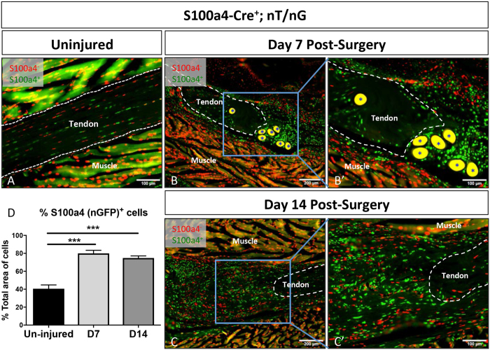
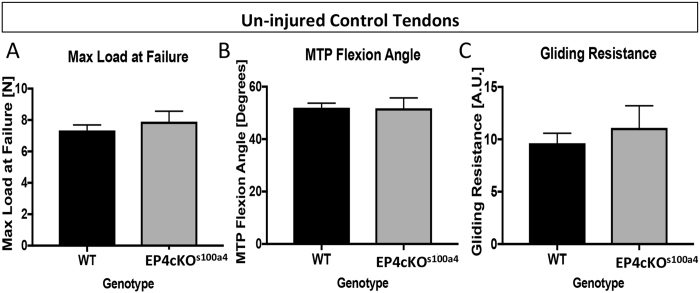
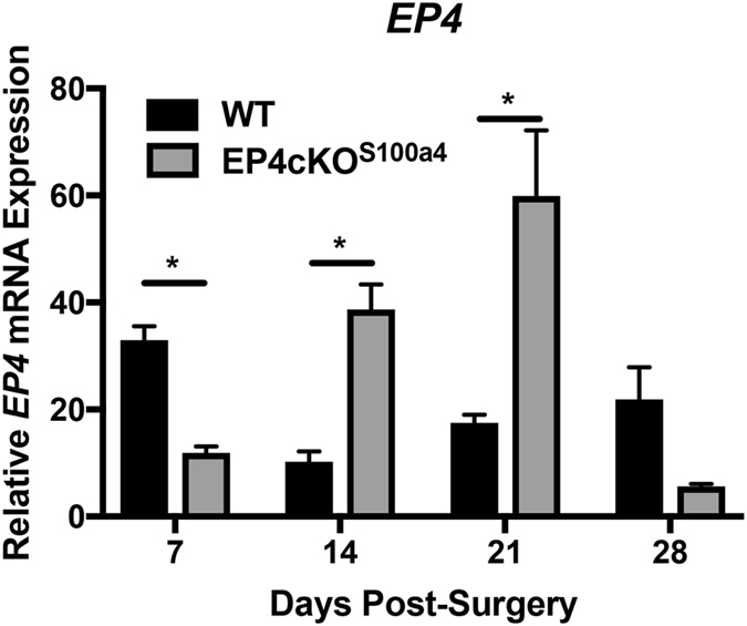
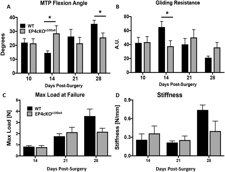
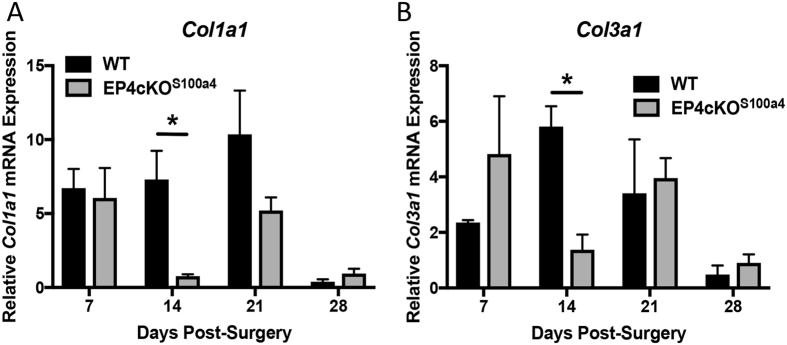
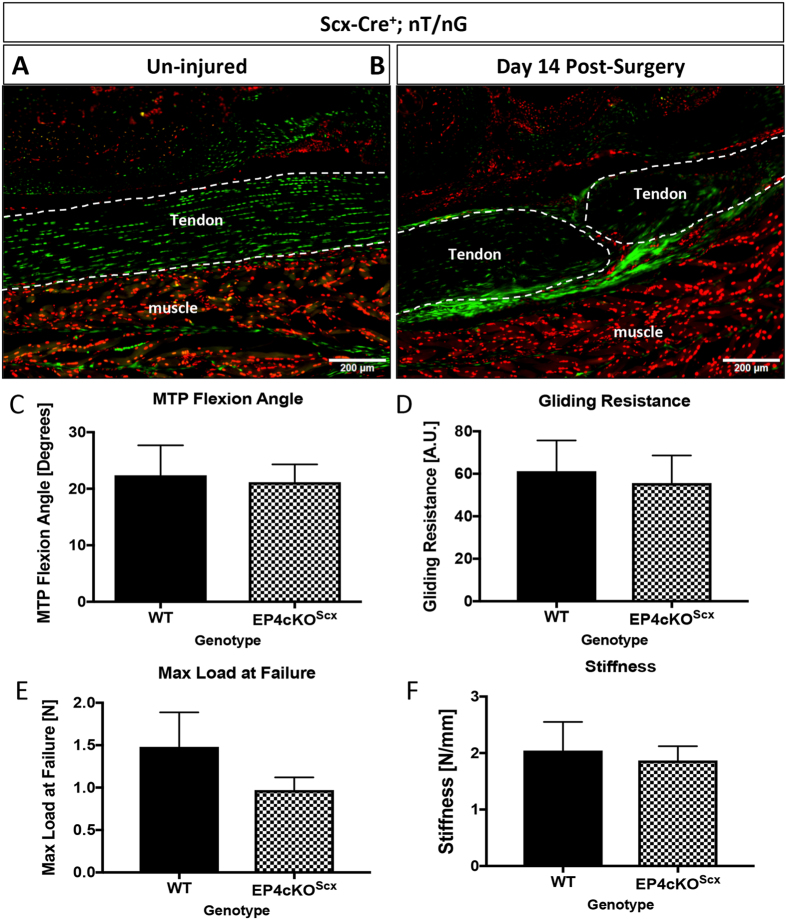
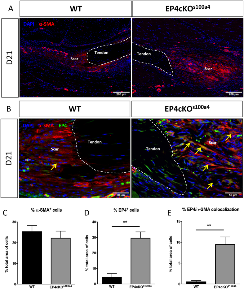
Tendon injuries heal via scar tissue rather than regeneration. This healing response forms adhesions between the flexor tendons in the hand and surrounding tissues, resulting in impaired range of motion and hand function. Mechanistically, inflammation has been strongly linked to adhesion formation, and Prostaglandin E2 (PGE2) is associated with both adhesion formation and tendinopathy. In the present study we tested the hypothesis that deletion of the PGE2 receptor EP4 in S100a4-lineage cells would decrease adhesion formation. S100a4-Cre; EP4 flox/flox (EP4cKOS100a4) repairs healed with improved gliding function at day 14, followed by impaired gliding at day 28, relative to wild type. Interestingly, EP4cKOS100a4 resulted in only transient deletion of EP4, suggesting up-regulation of EP4 in an alternative cell population in these mice. Loss of EP4 in Scleraxis-lineage cells did not alter gliding function, suggesting that Scx-lineage cells are not the predominant EP4 expressing population. In contrast, a dramatic increase in α-SMA+, EP4+ double-positive cells were observed in EP4cKOS100a4 suggesting that EP4cKOS100a4 repairs heal with increased infiltration of EP4 expressing α-SMA myofibroblasts, identifying a potential mechanism of late up-regulation of EP4 and impaired gliding function in EP4cKOS100a4 tendon repairs.


Conflict of interest statement

The authors declare that they have no competing interests.

Figures

Figure 1
Figure 1
S100a4-Cre effectively targets tendon. To demonstrate that S100a4-Cre effectively targets recombination in the tendon we examined healing in the double reporter nT/nG with S100a4-Cre+ cells expressing GFP, and S100a4-Cre cells expressing Td Tomato Red. S100a4-Cre results in recombination in both the (A) un-injured and (B & C) repaired tendon at days 7 (B & B’) and 14 (C & C’) post-surgery. Tendon tissue is outlined in white, (*) indicate auto-fluorescent sutures at the repair site. Scale bars (A,B’,C’) represent 100 microns, and (B & C) 200 microns. (D) Quantification of the proportion of nGFP+ (S100a4-Cre+) cells in the un-injured tendon and at 7 and 14 days post-repair. (***) Indicates p < 0.001 vs. un-injured.
Figure 2
Figure 2
EP4cKOS100a4 does not alter gliding function or mechanical properties of un-injured tendons. No changes in (A) Max load at failure, (B) MTP Flexion Angle, or (C) Gliding Resistance were observed between un-injured tendons from WT and EP4cKOS100a4 flexor tendons. For tensile testing, all un-injured tendons failed at the tendon mid-substance.
Figure 3
Figure 3
EP4cKOS100a4 significantly decreases EP4 expression during early healing. mRNA was isolated from EP4cKOS100a4 and WT tendon repairs between 7–28 days post-surgery. Relative EP4 expression was significantly decreased in EP4cKOS100a4 relative to WT at day 7, and was significantly increased on days 14 and 21 post-surgery. Expression was normalized to the internal control β-actin. (*) Indicates p < 0.05.
Figure 4
Figure 4
EP4cKOS100a4 significantly improves early gliding function without compromising mechanical properties during flexor tendon healing. (A) MTP Flexion Angle, and (B) Gliding Resistance were measured in WT and EP4cKOS100a4 flexor tendons between 10–28 days post-surgery. (C) Max load at failure, and (D) Stiffness were assessed between 14–28 days post-surgery. For tensile testing, all repaired tendons failed at the suture site. (*) Indicates p < 0.05 between WT and EPcKOS100a4 at the same time-point.
Figure 5
Figure 5
EP4cKOS100a4 repairs heal with decreased expression of Col1a1 and Col3a1. mRNA was isolated from EP4cKOS100a4 and WT mice between 7–28 days post-surgery. A significant decrease in both (A) Col1a1, and (B) Col3a1 expression was observed at day 14 post-surgery in EP4cKOS100a4 relative to WT. Expression was normalized to the internal control β-actin. (*) Indicates p < 0.05.
Figure 6
Figure 6
EP4cKOScx does not alter tendon healing. (A,B) Tracing of Scx-lineage cells using Scx-Cre; nT/nG demonstrates that Scx-lineage cells are present in the un-injured tendon (A), and the repaired tendon at day 14 post-surgery (B). Scale bar represents 100 microns. (CF) Repaired tendons from EP4cKOScx and WT mice were harvested at day 14 post-surgery. No changes in (C) MTP Flexion Angle, (D) Gliding Resistance, (E) Max load at failure, (F) Stiffness were observed between WT repairs and EP4cKOScx repairs. For tensile testing, all repaired tendons failed at the suture site.
Figure 7
Figure 7
EP4-expressing α-SMA myofibroblasts are increased in EP4cKOS100a4 repairs during later healing. (A) Abundant expression of α-SMA+ was observed in EP4cKOS100a4 and WT repairs on day 21 post-surgery. α-SMA+ cells are identified by red fluorescent staining, while nuclear stain DAPI is blue. Scale bars represent 200 microns. (B) Co-immunofluorescence for α-SMA (red) and EP4 (green) demonstrate very few α-SMA+, EP4+ cells (yellow arrow) in WT repairs at day 21. In contrast, abundant α-SMA+, EP4+ cells (yellow arrows) are observed in EP4cKOS100a4 repairs at this time. Scale bars represent 50 microns. (DF) Quantitative assessment of (D) α-SMA+ cells, (E) EP4+ cells, and (F) α-SMA, EP4 double positive cells by immunofluorescence at day 21. (**) Indicates p < 0.01 between WT and EP4cKOS100a4 repairs.

References

    1. Feldscher SB, Schneider LH. Flexor tenolysis. Hand Surg. 2002;7:61–74. doi: 10.1142/S0218810402000819. - DOI - PubMed
    1. Virchenko O, Skoglund B, Aspenberg P. Parecoxib impairs early tendon repair but improves later remodeling. Am J Sports Med. 2004;32:1743–1747. doi: 10.1177/0363546504263403. - DOI - PubMed
    1. Rouhani A, Tabrizi A, Ghavidel E. Effects of non-steroidal anti-inflammatory drugs on flexor tendon rehabilitation after repair. Arch Bone Jt Surg. 2013;1:28–30. - PMC - PubMed
    1. Khan MH, Li Z, Wang JH. Repeated exposure of tendon to prostaglandin-E2 leads to localized tendon degeneration. Clin J Sport Med. 2005;15:27–33. doi: 10.1097/00042752-200501000-00006. - DOI - PubMed
    1. Koshima H, et al. Expression of interleukin-1beta, cyclooxygenase-2, and prostaglandin E2 in a rotator cuff tear in rabbits. J Orthop Res. 2007;25:92–97. doi: 10.1002/jor.20241. - DOI - PubMed

Publication types

MeSH terms

Substances