Multicenter Study
. 2017 Aug 17;7(1):8671.
doi: 10.1038/s41598-017-07242-4.

Progress towards non-invasive diagnosis and follow-up of celiac disease in children; a prospective multicentre study to the usefulness of plasma I-FABP

Collaborators, Affiliations
Multicenter Study

Progress towards non-invasive diagnosis and follow-up of celiac disease in children; a prospective multicentre study to the usefulness of plasma I-FABP

M P M Adriaanse et al. Sci Rep. .

Abstract

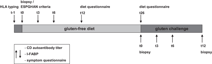
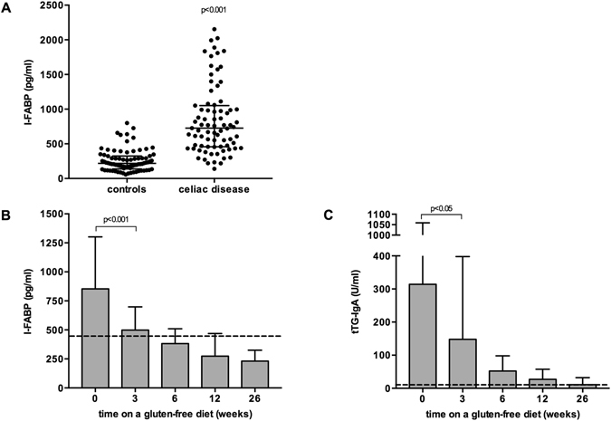
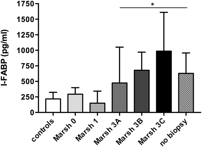
This prospective study investigates whether measurement of plasma intestinal-fatty acid binding protein (I-FABP), a sensitive marker for small intestinal epithelial damage, improves non-invasive diagnosing of celiac disease (CD), and whether I-FABP levels are useful to evaluate mucosal healing in patients on a gluten-free diet (GFD). Ninety children with elevated tTG-IgA titres and HLA-DQ2/DQ8 positivity were included (study group). Duodenal biopsies were taken, except in those fulfilling the ESPGHAN criteria. Plasma I-FABP levels and tTG-IgA titres were assessed sequentially during six months of follow-up. Eighty children with normal tTG-IgA titres served as control group. In 61/90 (67.8%) of the children in the study group an increased I-FABP level was found; in all these children CD diagnosis was confirmed. Interestingly, in 14/30 (46.7%) children with slightly elevated tTG-IgA titres (<10x upper limit of normal), an increased I-FABP level was found. In all these children the diagnosis of CD was confirmed histologically. After gluten elimination for six weeks I-FABP levels had decreased towards levels in the control group. Measurement of plasma I-FABP, in addition to tTG-IgA, EMA-IgA and HLAtyping, enables non-invasive diagnosing of CD in a substantial number of children, and might therefore be of value in the diagnostic approach of CD.


Conflict of interest statement

The authors declare that they have no competing interests.

Figures

Figure 1
Figure 1
Study protocol.
Figure 2
Figure 2
Study visits and measurements at disease presentation and during follow-up of all included patients.
Figure 3
Figure 3
Plasma I-FABP results of all included patients with a clinical suspicion of celiac disease.
Figure 4
Figure 4
Plasma I-FABP levels are significantly elevated in celiac disease patients compared to control individuals (A). Plasma I-FABP levels (B) and tTG-IgA titres (C) in celiac disease patients after initiation of a gluten-free diet (Figure B includes patients with elevated I-FABP levels at diagnosis only, n = 79, 21, 74, 70, 76 at t = 0, 3, 6, 12 and 26, respectively). *Significantly different as compared with week 0, p < 0.05.
Figure 5
Figure 5
Plasma I-FABP levels stratified for the degree of villous atrophy at time of diagnosis of CD. *Significantly elevated compared with controls, #Significantly elevated compared with Marsh 0, and Marsh I.
Figure 6
Figure 6
Correlation between plasma I-FABP levels and serum tTG-IgA titres at time of presentation.

References

    1. Green PH, Cellier C. Celiac disease. N. Engl. J. Med. 2007;357:1731–43. doi: 10.1056/NEJMra071600. - DOI - PubMed
    1. Husby S, et al. European Society for Pediatric Gastroenterology, Hepatology, and Nutrition guidelines for the diagnosis of coeliac disease. J. Pediatr. Gastroenterol. Nutr. 2012;54:136–60. doi: 10.1097/MPG.0b013e31821a23d0. - DOI - PubMed
    1. Mubarak A, Nikkels P, Houwen R, Ten Kate F. Reproducibility of the histological diagnosis of celiac disease. Scand. J. Gastroenterol. 2011;46:1065–73. doi: 10.3109/00365521.2011.589471. - DOI - PubMed
    1. Arguelles-Grande C, Tennyson CA, Lewis SK, Green PH, Bhagat G. Variability in small bowel histopathology reporting between different pathology practice settings: impact on the diagnosis of coeliac disease. J. Clin. Pathol. 2012;65:242–7. doi: 10.1136/jclinpath-2011-200372. - DOI - PubMed
    1. Ravelli A, et al. How patchy is patchy villous atrophy?: distribution pattern of histological lesions in the duodenum of children with celiac disease. Am. J. Gastroenterol. 2010;105:2103–10. doi: 10.1038/ajg.2010.153. - DOI - PubMed

Publication types

MeSH terms