. 2017 Oct 11;17(10):6235-6240.
doi: 10.1021/acs.nanolett.7b02929. Epub 2017 Sep 18.

Nanostructured Fibrous Membranes with Rose Spike-Like Architecture

Affiliations

Nanostructured Fibrous Membranes with Rose Spike-Like Architecture

Amir Nasajpour et al. Nano Lett. .

Abstract

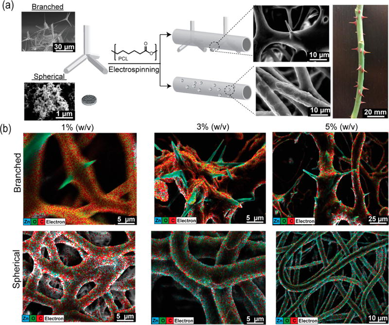
Nanoparticles have been used for engineering composite materials to improve the intrinsic properties and/or add functionalities to pristine polymers. The majority of the studies have focused on the incorporation of spherical nanoparticles within the composite fibers. Herein, we incorporate anisotropic branched-shaped zinc oxide (ZnO) nanoparticles into fibrous scaffolds fabricated by electrospinning. The addition of the branched particles resulted in their protrusion from fibers, mimicking the architecture of a rose stem. We demonstrated that the encapsulation of different-shape particles significantly influences the physicochemical and biological activities of the resultant composite scaffolds. In particular, the branched nanoparticles induced heterogeneous crystallization of the polymeric matrix and enhance the ultimate mechanical strain and strength. Moreover, the three-dimensional (3D) nature of the branched ZnO nanoparticles enhanced adhesion properties of the composite scaffolds to the tissues. In addition, the rose stem-like constructs offered excellent antibacterial activity, while supporting the growth of eukaryote cells.

Keywords: Branched tetrapod nanoparticles; antimicrobial; electrospinning; nanocomposites; scaffolds; zinc oxide.


Figures

Figure 1
Figure 1
Fabrication of the rose stem-like composite constructs containing spherical and branched nanoparticles. (a) Schematic illustration of fibrous composite fabrication: ZnO particles are dispersed within a PCL solution and extruded under high voltage. SEM images show the inability to confine the branched particle with consequent protrusion formation (rose stem-like structure). (b) EDAX map of dibranched and spherical ZnO particles at different concentrations (1, 3, and 5% w/v) and their distribution into the fibers. Elemental map demonstrates the distribution of Zn (blue), O (green), and C (red) in the composites.
Figure 2
Figure 2
Chemical and physical characterization of fibrous composite scaffolds. (a) Fourier transformer infrared spectroscopy (FTIR) spectra of the tested fibers. The inset within the graph highlights the carbonyl stretching peaks. (b) Wide-angle X-ray diffraction (WAXD) showing the effect of the ZnO nanoparticles on the carrier polymer (PCL) (110) and (200) crystal planes. The inset demonstrates that the incorporation of ZnO induces narrowing of the crystal planes, correlating with an enhanced crystallization, compared to the control (purple). (c) Two-dimensional WAXD images show the effect of ZnO nanoparticles on the reflection banding arc’s annotated within with arrows.
Figure 3
Figure 3
Mechanical and surface characterization of the nanocomposite structures. (a) Comparison of ultimate strength among the control sample (PCL), branched, and spherical composite scaffolds. The reports of statistical analysis placed directly on top of the bars refer to the comparison of the sample with the control (pristine PCL). The comparison between different samples is shown by lines. (b) Comparison of constructs’ Young’s modulus. (c) Representative lap shear curves of scaffolds sandwiched between porcine skins. (d) Comparison of the adhesion strength of different composite structures (data is derived from lap shear tests with porcine skin). (e) Representative SEM images from top and cross-sectional view of a branched composite material after failure during lap shear test. (f) Measurements of contact angle between a deionized water drop and the material surfaces. (g) Schematic illustration of fibrous structures wetting behavior (top). coupled with optical imaging analysis (bottom) (n = 4 for ultimate strength and Young’s modulus data, n = 3 for adhesion strength data, and n = 6 for contact angle data, *: P < 0.33, **: P < 0.002, ***: P < 0.001).
Figure 4
Figure 4
Characterization of the antimicrobial and biological properties of the composite scaffolds. (a) Representative SEM images of control (PCL), branched, and spherical samples after 24 h incubation with E. coli (top) and P. aeruginosa. (bottom) (b) Number of colony forming units (CFU) over the samples after 24 h incubation with E. coli (top) and P. aeruginosa (bottom). The arrows are showing the presence of bacteria on the surface of the nanofibers. (c) Representative SEM images of keratinocytes attachment to control, branched, and spherical composite scaffolds. (d) Metabolic activity of HACATs assessed by PrestoBlue assay after 1 and 3 days of culture over pristine PCL compared with ZnO nanocomposites with different concentrations (1, 3, 5%) of branched and spherical ZnO nanoparticles. (n = 7 for antimicrobial tests, n = 5 for metabolic activity tests, *: P < 0.33, **: P < 0.002, ***: P < 0.001).

References

    1. Leijten J, Rouwkema J, Zhang YS, Nasajpour A, Dokmeci MR, Khademhosseini A. Small. 2016;12:2130–2145. - PMC - PubMed
    1. Li D, Xia Y. Adv. Mater. 2004;16:1151–1170.
    1. Persano L, Dagdeviren C, Su Y, Zhang Y, Girardo S, Pisignano D, Huang Y, Rogers JA. Nat. Commun. 2013;4:1633. - PubMed
    1. Najafabadi AH, Tamayol A, Annabi N, Ochoa M, Mostafalu P, Akbari M, Nikkhah M, Rahimi R, Dokmeci MR, Sonkusale S, Ziaie B, Khademhosseini A. Adv. Mater. 2014;26:5823–5830. - PMC - PubMed
    1. Goldberg M, Langer R, Jia X. J. Biomater. Sci., Polym. Ed. 2007;18:241–268. - PMC - PubMed

Publication types

MeSH terms