. 2017 Sep;31(9):1308-1316.
doi: 10.1038/eye.2017.158. Epub 2017 Aug 18.

Technique of retinal gene therapy: delivery of viral vector into the subretinal space

Affiliations

Technique of retinal gene therapy: delivery of viral vector into the subretinal space

K Xue et al. Eye (Lond). 2017 Sep.

Abstract

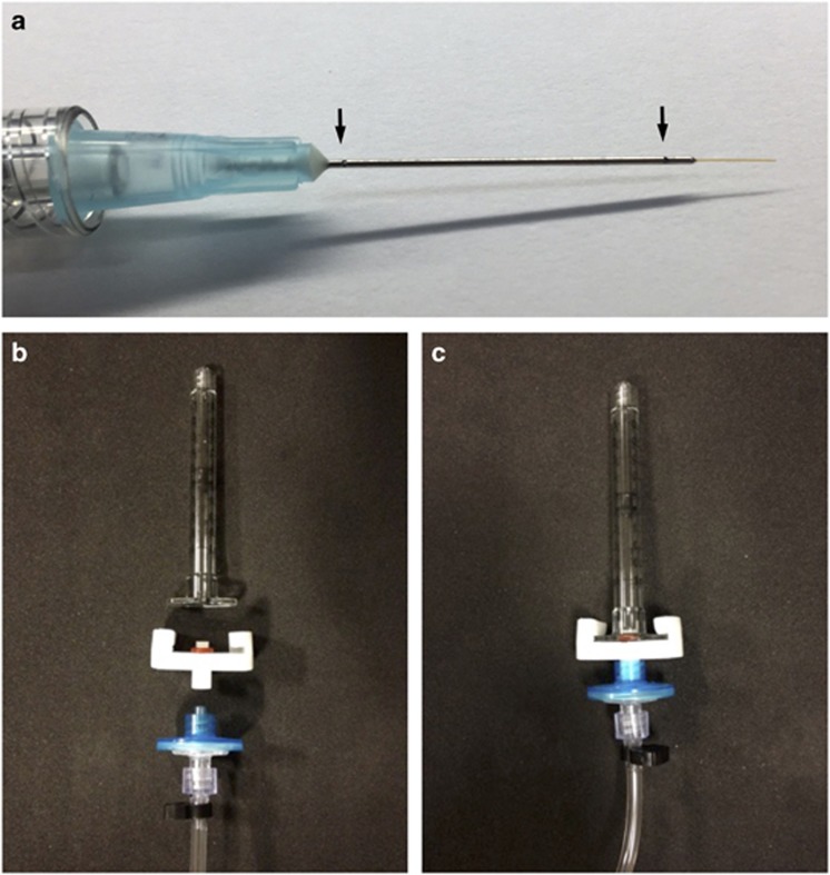
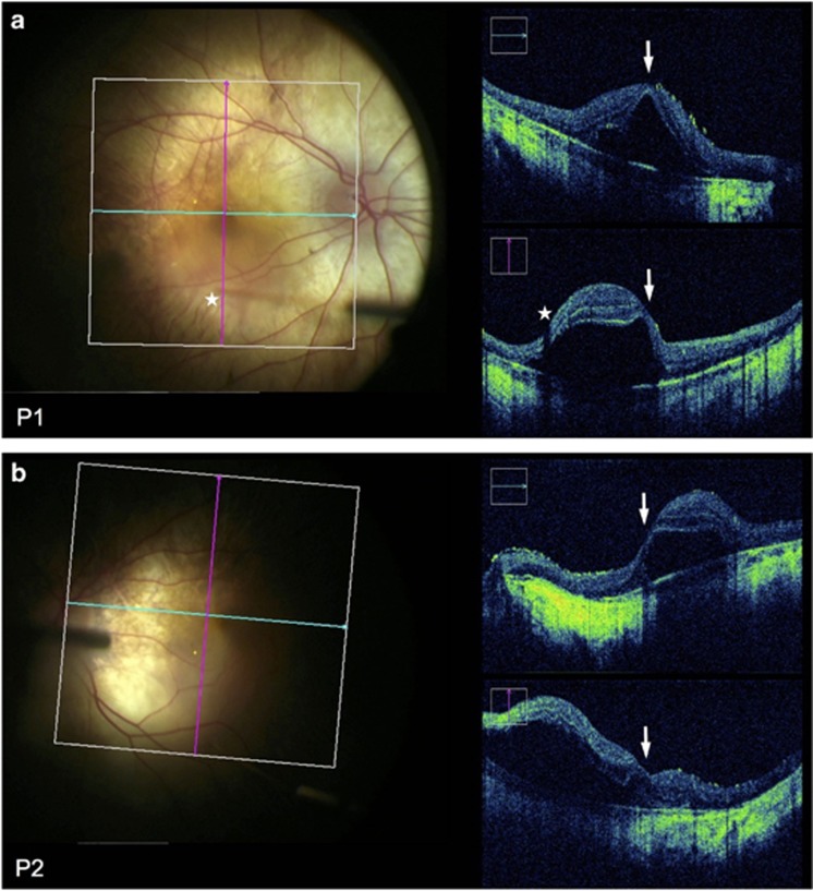
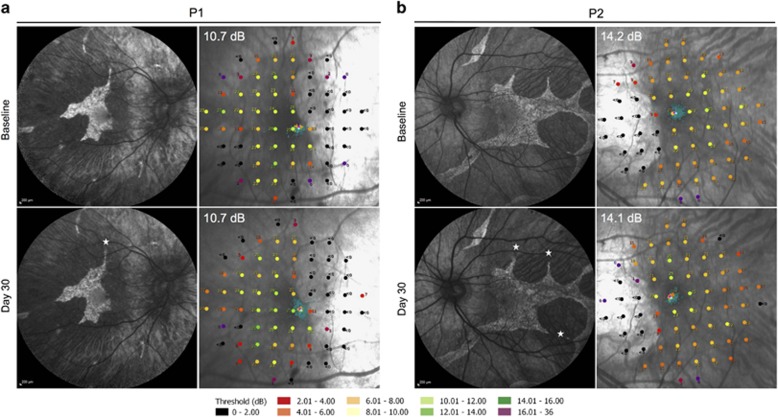
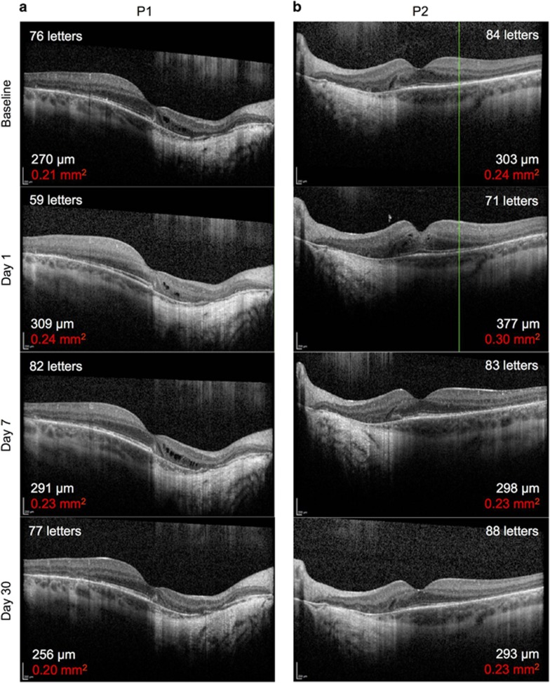
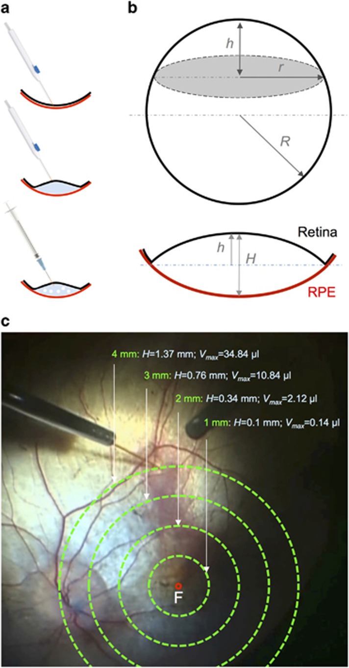
PurposeSafe and reproducible delivery of gene therapy vector into the subretinal space is essential for successful targeting of the retinal pigment epithelium (RPE) and photoreceptors. The success of surgery is critical for the clinical efficacy of retinal gene therapy. Iatrogenic detachment of the degenerate (often adherent) retina in patients with hereditary retinal degenerations and small volume (eg, 0.1 ml) subretinal injections pose new surgical challenges.MethodsOur subretinal gene therapy technique involved pre-operative planning with optical coherence tomography (OCT) and autofluorescence (AF) imaging, 23 G pars plana vitrectomy, internal limiting membrane staining with Membrane Blue Dual (DORC BV, Zuidland, Netherlands), a two-step subretinal injection using a 41 G Teflon tipped cannula (DORC) first with normal saline to create a parafoveal bleb followed by slow infusion of viral vector via the same self-sealing retinotomy. Surgical precision was further enhanced by intraoperative OCT (Zeiss Rescan 7000, Carl Zeiss Meditec AG, Jena, Germany). Foveal functional and structural recovery was evaluated using best-corrected Early Treatment Diabetic Retinopathy Study (ETDRS) visual acuity, microperimetry and OCT.ResultsTwo patients with choroideremia aged 29 (P1) and 27 (P2) years, who had normal and symmetrical levels of best-corrected visual acuity (BCVA) in both eyes, underwent unilateral gene therapy with the fellow eye acting as internal control. The surgeries were uncomplicated in both cases with successful detachment of the macula by subretinal vector injection. Both treated eyes showed recovery of BCVA (P1: 76-77 letters; P2: 84-88 letters) and mean threshold sensitivity of the central macula (P1: 10.7-10.7 dB; P2: 14.2-14.1 dB) to baseline within a month. This was accompanied by normalisation of central retinal thickness on OCT.ConclusionsHerein we describe a reliable technique for subretinal gene therapy, which is currently used in clinical trials to treat choroideremia using an adeno-associated viral (AAV) vector encoding the CHM gene. Strategies to minimise potential complications, such as avoidance of excessive retinal stretch, air bubbles within the injection system, reflux of viral vector and post-operative vitritis are discussed.


Conflict of interest statement

REM: a founding director of NightstaRx (London, UK)—a gene therapy company established by the University of Oxford and funded by the Wellcome Trust. REM is a named inventor on a patent ‘Vector administration and dosing’ filed on behalf of the University of Oxford (US Patent Application No. 14/598,948). The remaining authors declare no conflict of interest.

Figures

Figure 1
Figure 1
Subretinal gene therapy injection system. (a) A dual-bore 41 G blunt polytetrafluoroethylene (Teflon) tipped cannula mounted within a 23 G steel shaft (DORC, Zuidland, The Netherlands). Arrows indicate proximal and distal pressure relief holes along the shaft, which allow evacuation of excess vitreous fluid out of the eye during the injection. (b) Components of the subretinal injection system consisting of a 1 ml BD syringe, custom syringe lock with a rubber O-ring, air filter and polyethylene connection tubing (Alchimia, Ponte S Nicolò, Italy). (c) Assembled injection system.
Figure 2
Figure 2
OCT-guided subretinal injection. Microscope photograph and simultaneous intraoperative OCT (Zeiss Rescan 7000, Carl Zeiss Meditec AG, Jena, Germany) during subretinal gene therapy for choroideremia in Patient 1 OD (a) and Patient 2 OS (b). Blue and pink grid lines indicate the locations of horizontal and vertical OCT B-scans respectively. The fovea could be seen to be detached in both cases (arrows). Stars indicate the tip of the subretinal cannula (seen out of focus) and the corresponding shadow cast on the OCT.
Figure 3
Figure 3
Preservation of autofluorescence (AF) island and central retinal sensitivity following subretinal gene therapy. Baseline and 30 days post-operative AF images and 10-2 MAIA microperimetry of the treated eye of Patient 1 (a) and Patient 2 (b) are shown. Stars indicate the locations of retinotomies (one in P1 and three in P2) performed during the subretinal BSS and vector injections, which successfully detached the central retinal island in each case. The threshold retinal sensitivities of the central 20° of the macula were represented by 68 colour-coded points superimposed on the cSLO image. The points of fixation are shown as clusters of fine green dots.
Figure 4
Figure 4
Resolution of subretinal fluid and recovery of visual acuity following subretinal gene therapy. Baseline and post-operative day 1, 7 and 30 foveal OCT images from Patient 1 (a) and Patient 2 (b) as described in Figures 2 and 3. The best-correct visual acuity (number of ETDRS letters) at each time-point is shown in the top corner of each OCT image. The mean central retinal thickness of the 1 mm2 ETDRS circle (in μm) and retinal volume from the ILM to the Bruch’s membrane within the 1 mm2 ETDRS circle (in mm2, red) are shown in the bottom corner of each OCT image. No significant changes in mean central retinal thickness or volume were observed in the fellow eye of either patient (Supplementary Table I).
Figure 5
Figure 5
The principle of subretinal bleb formation during gene therapy for choroideremia. (a) A schematic drawing of key surgical steps consisting of the initiation of retinal detachment by subretinal injection of basic saline solution (BSS) using an extendible 41 G cannula (DORC), a necessary step to overcome the abnormally adherent retina of patients with outer retinal degenerations. The AAV vector solution is then loaded into a 1 ml syringe and slowly infused into the subretinal bleb through the same retinotomy using a dual-bore 41 G cannula (DORC). (b) The spherical cap formula can be used to calculate the height of the subretinal bleb (H=2 h) in relation to the distance of the retinotomy from the fovea (r) and the radius of the whole eye (R, assumed to be half of the average axial length of 24 mm). (c) If the retina was assumed to ‘flip’ from concave to convex without undergoing any elastic stretch during a subretinal injection, for each diameter of subretinal bleb (r=1, 2, 3 or 4 mm), the maximum bleb volume (Vmax) was calculated to be 0.14, 2.12, 10.84 and 34.84 μl, respectively.

References

    1. Jacobson SG, Cideciyan AV, Roman AJ, Sumaroka A, Schwartz SB, Heon E et al. Improvement and decline in vision with gene therapy in childhood blindness. N Engl J Med 2015; 372: 1920–1926. - PMC - PubMed
    1. Bainbridge JW, Mehat MS, Sundaram V, Robbie SJ, Barker SE, Ripamonti C et al. Long-term effect of gene therapy on Leber's congenital amaurosis. N Engl J Med 2015; 372: 1887–1897. - PMC - PubMed
    1. Edwards TL, Jolly JK, Groppe M, Barnard AR, Cottriall CL, Tolmachova T et al. Visual Acuity after Retinal Gene Therapy for Choroideremia. N Engl J Med 2016; 374: 1996–1998. - PMC - PubMed
    1. Bennett J, Wellman J, Marshall KA, McCague S, Ashtari M, DiStefano-Pappas J et al. Safety and durability of effect of contralateral-eye administration of AAV2 gene therapy in patients with childhood-onset blindness caused by RPE65 mutations: a follow-on phase 1 trial. Lancet 2016; 388: 661–672. - PMC - PubMed
    1. Ghazi NG, Abboud EB, Nowilaty SR, Alkuraya H, Alhommadi A, Cai H et al. Treatment of retinitis pigmentosa due to MERTK mutations by ocular subretinal injection of adeno-associated virus gene vector: results of a phase I trial. Hum Genet 2016; 135: 327–343. - PubMed

Substances