. 2017 Oct 3;13(10):1754-1766.
doi: 10.1080/15548627.2017.1357792. Epub 2017 Aug 18.

BNIP3L/NIX-mediated mitophagy protects against ischemic brain injury independent of PARK2

Affiliations

BNIP3L/NIX-mediated mitophagy protects against ischemic brain injury independent of PARK2

Yang Yuan et al. Autophagy. .

Abstract

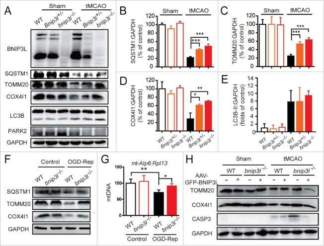
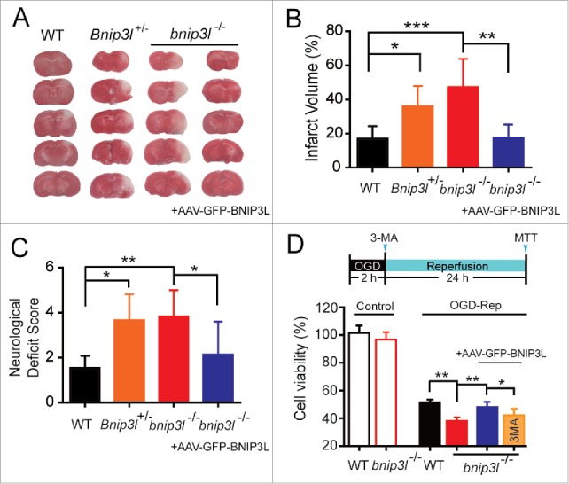
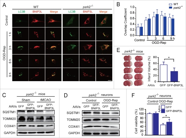
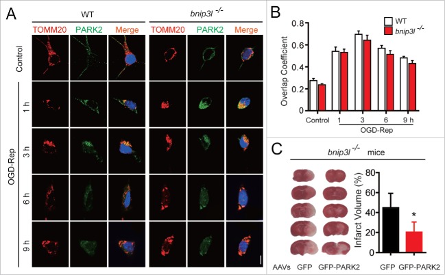
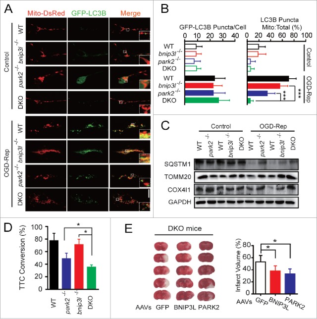
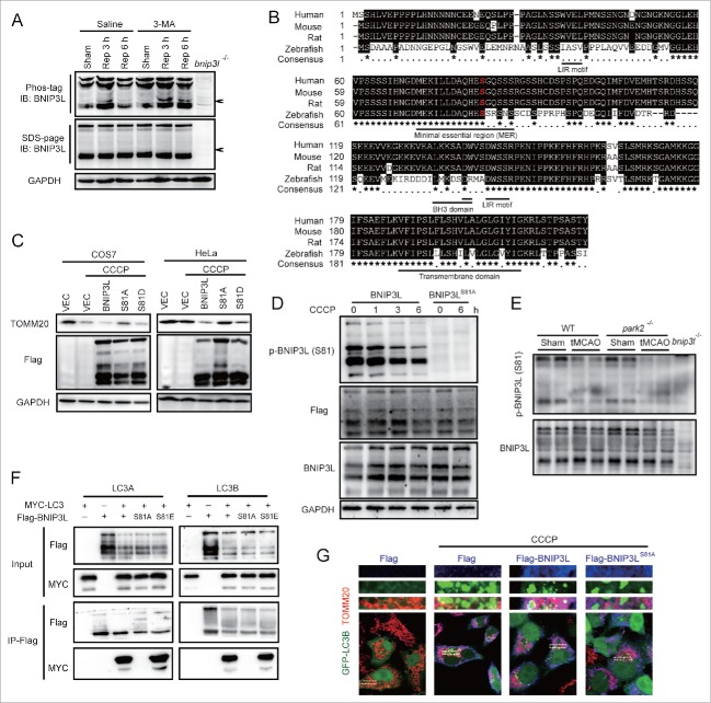
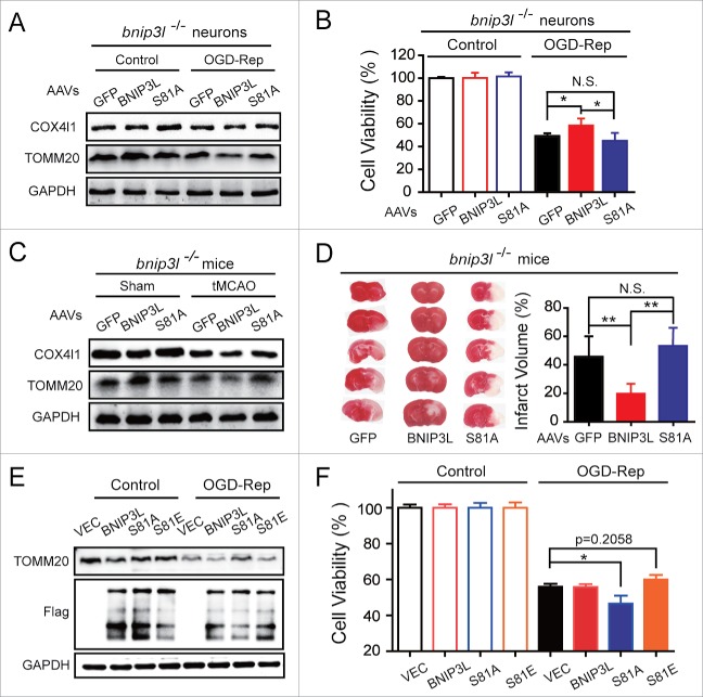
Cerebral ischemia induces massive mitochondrial damage. These damaged mitochondria are cleared, thus attenuating brain injury, by mitophagy. Here, we identified the involvement of BNIP3L/NIX in cerebral ischemia-reperfusion (I-R)-induced mitophagy. Bnip3l knockout (bnip3l-/-) impaired mitophagy and aggravated cerebral I-R injury in mice, which can be rescued by BNIP3L overexpression. The rescuing effects of BNIP3L overexpression can be observed in park2-/- mice, which showed mitophagy deficiency after I-R. Interestingly, bnip3l and park2 double-knockout mice showed a synergistic mitophagy deficiency with I-R treatment, which further highlighted the roles of BNIP3L-mediated mitophagy as being independent from PARK2. Further experiments indicated that phosphorylation of BNIP3L serine 81 is critical for BNIP3L-mediated mitophagy. Nonphosphorylatable mutant BNIP3LS81A failed to counteract both mitophagy impairment and neuroprotective effects in bnip3l-/- mice. Our findings offer insights into mitochondrial quality control in ischemic stroke and bring forth the concept that BNIP3L could be a potential therapeutic target for ischemic stroke, beyond its accepted role in reticulocyte maturation.

Keywords: BNIP3L/NIX; PARK2/PARKIN; cerebral ischemia; mitophagy; phosphorylation.


Figures

Figure 1.
Figure 1.
BNIP3L is required in ischemia-reperfusion-induced mitophagy. (A) Bnip3l+/+, Bnip3l+/−, and bnip3l/− mice were subjected to transient middle cerebral artery occlusion (tMCAO) for 1 h, and expression of SQSTM1, TOMM20, COX4I1, and LC3B in ischemic penumbra was assessed by western blot after 6 h of reperfusion. (B–E) Semi-quantitative analysis of SQSTM1, TOMM20, COX4I1, and LC3B bands are shown, respectively. (F and G) Primary cultured mice cortical neurons were subjected to 2 h of oxygen and glucose deprivation followed by 6 h of reperfusion (OGD-Rep). (F) The SQSTM1, TOMM20, and COX4I1 protein levels in both Bnip3l+/+ and bnip3l/− neurons treated with OGD-Rep were determined. (G) The relative mitochondrial DNA (mtDNA) levels, which as indicated by the mt-Atp6 (mitochondria-encoded DNA):Rpl13 (nucleus-encoded DNA) ratio, were assessed by real-time PCR in both Bnip3l+/+ and bnip3l/− neurons. (H) Adeno-associated viruses expressing GFP-BNIP3L or GFP were intracerebrally injected into Bnip3l+/+ and bnip3l/− mice 2 wk before tMCAO. After 6 h of reperfusion, the TOMM20, COX4I1, and cleaved-CASP3 expression in ischemic penumbra were determined by western blot. The data are expressed as means ± SD. Statistical comparisons were performed with one-way ANOVA followed by Dunnett's t test. *p < 0.05, **p < 0.01, ***p < 0.001 vs. the indicated group.
Figure 2.
Figure 2.
BNIP3L deficiency aggravates ischemic brain injury. (A) Mice were subjected to 1 h of tMCAO followed by 24 h reperfusion. The cerebral infarct volumes were determined by TTC staining; representative TTC-stained brain slices from each group are shown. (B and C) The infarct volumes and neurological deficit scores of each group were determined (n = 5–8). (D) A timeline scheme showed the procedures of this experiment. Primary cultured bnip3l/− neurons were infected with adeno-associated viruses expressing GFP-BNIP3L or GFP 48 h before 2 h of oxygen and glucose deprivation treatment. 3-MA (10 μg) was injected intracerebroventricularly at the onset of reperfusion. After 24 h of reperfusion, the cell viability in both Bnip3l+/+ and bnip3l/− neurons was measured by MTT assay. The data are expressed as means ± SD. Statistical comparisons were performed with one-way ANOVA followed by Dunnett's t test. *p < 0.05, **p< 0.01, ***p < 0.001 vs. the indicated group.
Figure 3.
Figure 3.
PARK2 is dispensable in BNIP3L-mediated mitophagy evoked by ischemia-reperfusion. (A) Park2+/+ and park2/− neurons were treated with 2 h of OGD. LC3B (green) and BNIP3L (red) were stained by immunocytochemistry at the indicated time after reperfusion, and the images were taken by confocal microscopy. (B) Columns represent the Manders overlap coefficient of BNIP3L and LC3B in Park2+/+and park2−/− neurons. At least 30 cells from 3 independent experiments for each group were included. (C and D) park2/− mice and primary cultured neurons were infected with AAVs expressing GFP-BNIP3L or GFP. The mice were subjected to one h of tMCAO with 6 h of reperfusion. The cultured neurons were subjected to 2 h of OGD and 6 h of reperfusion. The SQSTM1, TOMM20 and COX4I1 levels in (C) brain tissue and (D) cultured neurons were determined by western blot. (E) Another set of mice were killed 24 h after MCAO and infarct volumes were determined by TTC staining. (F) Primary cultured wild-type and park2/− neurons were infected with GFP-BNIP3L or GFP-expressing AAVs 48 h in advance. After 2 h of OGD and 24 h of reperfusion, the cell viability was measured by MTT assay. The data are expressed as means ± SD. Statistical comparisons were performed with unpaired Student t tests. *p < 0.05 vs. the indicated group.
Figure 4.
Figure 4.
BNIP3L is dispensable in PARK2-dependent mitophagy evoked by ischemia-reperfusion. (A) Bnip3l+/+ and bnip3l/− neurons were treated with 2 h of OGD. DAPI (blue), PARK2 (green), and the mitochondrial marker TOMM20 (red) were stained by immunocytochemistry at the indicated times after reperfusion. The images were taken by confocal microscopy. (B) Columns represent the Manders overlap coefficient of PARK2 and TOMM20 in Bnip3l+/+ and bnip3l/− neurons. At least 44 cells from 3 independent experiments for each group were included. (C) bnip3l/− mice were infected with GFP- or GFP-PARK2-expressing AAVs, and were then subjected to tMCAO for one h. The mice were killed 24 h after MCAO, and infarct volumes were determined by TTC staining. *p < 0.05 vs. the indicated group. The data are expressed as means ± SD. Statistical comparisons were performed with one-way ANOVA followed by Dunnett's t test. *p < 0.05 vs. the indicated group.
Figure 5.
Figure 5.
Mitophagy is further impaired in bnip3l and park2 double-knockout ischemic brain. park2 and bnip3l double-knockout mice (DKO) were generated by hybridizing park2/− with bnip3l/− mice. (A) Primary cultured cortical neurons from the indicated germline mice were previously transfected with GFP-LC3B and Mito-DsRed by viral vector infection. After 3 h of reperfusion, fluorescent images were captured by confocal microscopy. Images show representative examples from 3 independent experiments. (B) Columns represent the number of GFP-LC3B-positive puncta per cell (left panel) and the proportion of LC3B puncta on mitochondria of the total LC3B puncta (right panel). At least 5 random fields from one section and 3 to 6 sections were averaged in each independent experiment. The bar chart shows mean ± SD values of puncta number from at least 3 independent experiments; at least 50 cells were analyzed in each group. (C and D) Brain corticostriatal slices from the indicated mice were subjected to 15 min of OGD plus 1 h reperfusion. (C) The protein levels of SQSTM1, TOMM20, and COX4I1 in the indicated mice were assessed by western blot. (D) After one h of reperfusion, tissue viability was quantified by TTC conversion assay. (E) DKO mice were injected with AAVs expressing GFP, GFP-BNIP3L or GFP-PARK2. The mice were then subjected to one h of tMCAO with 24 h of reperfusion. The representative images of ischemic brains are shown and the infarct volumes were determined by TTC staining. The data are expressed as means ± SD. Statistical comparisons were performed with one-way ANOVA followed by Dunnett's t test. *p < 0.05, **p < 0.01, ***p < 0.001 vs. the indicated group.
Figure 6.
Figure 6.
Phosphorylation of BNIP3L (Ser81) is responsible for BNIP3L-mediated mitophagy. (A) Wild-type mice were subjected to one h tMCAO, 10 µg 3-MA was injected at the onset of reperfusion, and phosphorylation of BNIP3L was determined by Phos-tag analysis at the indicated reperfusion time. (B) Alignment of BNIP3L sequences from the indicated species. Identical amino acids residues are highlighted in black. Highly conserved residues are labeled by “*” while less conserved ones are labeled by “•." Serine 81 is marked in red. Indicated domains of BNIP3L are underlined. (C) Plasmids expressing Flag (vector, VEC), Flag-BNIP3L, Flag-BNIP3LS81A and Flag-BNIP3LS81D were transfected into COS7 and HeLa cells for 36 h, and 12 h after 20 µM CCCP treatment the TOMM20 level was assessed by western blot. (D) Anti-phosphorylated BNIP3L (Ser81) antibody was confirmed in HeLa cells expressing Flag-BNIP3L or Flag-BNIP3LS81A after 20 µM CCCP treatment of the indicated hours. (E) The phosphorylation of BNIP3L on Ser81 was detected in both Park2+/+ and park2/− mice after tMCAO, sample from bnip3l−/− mice were taken as a negative control. (F) Protein-protein interactions of both LC3A and LC3B with BNIP3L, BNIP3LS81A and BNIP3LS81E were confirmed by co-immunoprecipitation in HeLa cells expressing the indicated plasmids. (G) Plasmids expressing Flag, Flag-BNIP3L and Flag-BNIP3LS81A were co-transfected with GFP-LC3B plasmid to HeLa cells for 36 h. Six hours after 20 µM CCCP treatment, Flag (blue) and the mitochondrial marker TOMM20 (red) were stained by immunocytochemistry and the images were taken by confocal microscopy.
Figure 7.
Figure 7.
Phosphorylation of BNIP3L on Ser81 is required for the protection of mitophagy in ischemia-reperfusion. (A and B) bnip3l/− neurons were transfected with adeno-associated viruses (AAVs) expressing GFP, GFP-BNIP3L, or GFP-BNIP3LS81A. (A) After 2 h of OGD followed with 6 h of reperfusion, the TOMM20 and COX4I1 protein levels were assessed by western blot. (B) After 24 h of reperfusion, the neuronal viability was measured by MTT assay. (C and D) bnip3l/− mice brains were infected with AAVs expressing GFP, GFP-BNIP3L, or GFP-BNIP3LS81A and were then subjected to one h of tMCAO. (C) After 6 h of reperfusion, TOMM20 and COX4I1 levels were assessed by western blot. (D) After 24 h of reperfusion, the infarct volumes were determined by TTC staining. (E and F) Wild-type neurons were electroporated with plasmids expressing Flag (vector, VEC), Flag-BNIP3L, Flag-BNIP3LS81A or Flag-BNIP3LS81E. (E) After 2 h of OGD followed with 6 h of reperfusion, the TOMM20 and Flag-BNIP3L proteins were assessed by western blot. (F) After 24 h of reperfusion, the neuronal viability was measured by MTT assay. The data are expressed as means ± SD. Statistical comparisons were performed with one-way ANOVA followed by Dunnett's t test. *p < 0.05, **p < 0.01 vs. the indicated group.

References

    1. Zhang X, Yan H, Yuan Y, Gao J, Shen Z, Cheng Y, Shen Y, Wang RR, Wang X, Hu WW, et al.. Cerebral ischemia-reperfusion-induced autophagy protects against neuronal injury by mitochondrial clearance. Autophagy. 2013;9:1321-33. doi:10.4161/auto.25132. PMID:23800795 - DOI - PubMed
    1. Yu S, Zheng S, Leng J, Wang S, Zhao T, Liu J. Inhibition of mitochondrial calcium uniporter protects neurocytes from ischemia/reperfusion injury via the inhibition of excessive mitophagy. Neurosci Letters. 2016;628:24-9. doi:10.1016/j.neulet.2016.06.012 - DOI - PubMed
    1. Zuo W, Zhang S, Xia CY, Guo XF, He WB, Chen NH. Mitochondria autophagy is induced after hypoxic/ischemic stress in a Drp1 dependent manner: the role of inhibition of Drp1 in ischemic brain damage. Neuropharmacology. 2014;86:103-15. doi:10.1016/j.neuropharm.2014.07.002. PMID:25018043 - DOI - PubMed
    1. Li Q, Zhang T, Wang J, Zhang Z, Zhai Y, Yang GY, Sun X. Rapamycin attenuates mitochondrial dysfunction via activation of mitophagy in experimental ischemic stroke. Biochem Biophys Res Commun. 2014;444:182-8. doi:10.1016/j.bbrc.2014.01.032. PMID:24440703 - DOI - PubMed
    1. Zhang X, Yuan Y, Jiang L, Zhang J, Gao J, Shen Z, Zheng Y, Deng T, Yan H, Li W, et al.. Endoplasmic reticulum stress induced by tunicamycin and thapsigargin protects against transient ischemic brain injury: Involvement of PARK2-dependent mitophagy. Autophagy 2014;10:1801-13. doi:10.4161/auto.32136. PMID:25126734 - DOI - PMC - PubMed

MeSH terms