

Autophagy-monitoring and autophagy-deficient mice
- PMID: 28820286
- PMCID: PMC5640176
- DOI: 10.1080/15548627.2017.1343770
Autophagy-monitoring and autophagy-deficient mice
Abstract
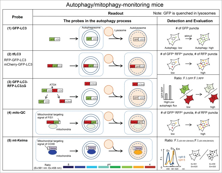
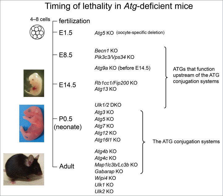
Discovery of yeast autophagy-related (ATG) genes and subsequent identification of their homologs in other organisms have enabled researchers to investigate physiological functions of macroautophagy/autophagy using genetic techniques. Specific identification of autophagy-related structures is important to evaluate autophagic activity, and specific ablation of autophagy-related genes is a critical means to determine the requirements of autophagy. Here, we review currently available mouse models, particularly focusing on autophagy (and mitophagy) indicator models and systemic autophagy-related gene-knockout mouse models.
Keywords: autophagy; knockout mouse; mitophagy; reporter mouse; selective autophagy.
Figures
References
-
- Mizushima N, Komatsu M. Autophagy: Renovation of cells and tissues. Cell. 2011;147:728-41. https://doi.org/ 10.1016/j.cell.2011.10.026. PMID:22078875. - DOI - PubMed
-
- Kabeya Y, Mizushima N, Ueno T, Yamamoto A, Kirisako T, Noda T, Kominami E, Ohsumi Y, Yoshimori T. LC3, a mammalian homologue of yeast Apg8p, is localized in autophagosome membranes after processing. EMBO J. 2000;19:5720-8. https://doi.org/ 10.1093/emboj/19.21.5720. PMID:11060023. - DOI - PMC - PubMed
-
- Mizushima N, Yamamoto A, Matsui M, Yoshimori T, Ohsumi Y. In vivo analysis of autophagy in response to nutrient starvation using transgenic mice expressing a fluorescent autophagosome marker. Mol Biol Cell. 2004;15:1101-11. https://doi.org/ 10.1091/mbc.E03-09-0704. PMID:14699058. - DOI - PMC - PubMed
-
- Kuma A, Hatano M, Matsui M, Yamamoto A, Nakaya H, Yoshimori T, Ohsumi Y, Tokuhisa T, Mizushima N. The role of autophagy during the early neonatal starvation period. Nature. 2004;432:1032-6. https://doi.org/ 10.1038/nature03029. PMID:15525940. - DOI - PubMed
-
- Vodicka P, Lim J, Williams DT, Kegel KB, Chase K, Park H, Marchionini D, Wilkinson S, Mead T, Birch H, et al. Assessment of chloroquine treatment for modulating autophagy flux in brain of WT and HD mice. J Huntingtons Dis. 2014;3:159-74. https://doi.org/ 10.4161/auto.4451. PMID:25062859. - DOI - PubMed
Publication types
MeSH terms
Substances
LinkOut - more resources
Full Text Sources
Other Literature Sources