. 2017 Sep;97(3):767-770.
doi: 10.4269/ajtmh.16-0722. Epub 2017 Aug 18.

The Phenotype of Circulating Neutrophils during Visceral Leishmaniasis

Affiliations

The Phenotype of Circulating Neutrophils during Visceral Leishmaniasis

Smriti Sharma et al. Am J Trop Med Hyg. 2017 Sep.

Abstract

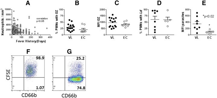
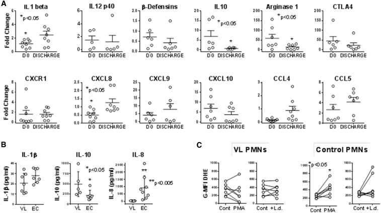
Visceral leishmaniasis (VL) is a chronic parasitic disease associated with suppressed T cell responses. Although parasites reside intracellularly in macrophages during chronic VL, neutrophils are the first host cell to infiltrate the infection site and phagocytose the parasite. Subsets of neutrophils with unusual characteristics have been documented in human VL, but whether the total neutrophil population is aberrant during disease is not known. Therefore, we examined phenotypic characteristics of unfractionated polymorphonuclear leukocyte (neutrophils) from subjects with active VL, and compared these with neutrophils from healthy controls or subjects who have been treated for VL. The data showed decreased mRNA and diminished amounts of the neutrophil chemoattractant CXCL8 (interleukin [IL]-8), increased IL-10 mRNA and protein, and elevated transcripts encoding arginase-1, which is involved in suppressing T cell responses. Neutrophils from VL subjects showed enhanced capacity to phagocytose Leishmania spp. promastigotes. The results suggest that neutrophils may contribute to immunosuppression in subjects with active VL.


Figures

Figure 1.
Figure 1.
Neutrophil abundance and phagocytic capacity. (A) Peripheral blood neutrophil counts from subjects with visceral leishmaniasis (VL) were plotted against the number of days of fever according to the patient history. Data were analyzed with Spearman nonparametric correlation. (BE) polymorphonuclear leukocyte (neutrophils) (PMNs) were isolated from peripheral blood of subjects with acute VL or from endemic controls (EC). PMNs were incubated with either FITC-labeled opsonized zymosan (OZ) or opsonized Leishmania donovani promastigotes for 30 minutes. The percentage of CD66b + cells with fluorescence indicating associated OZ (B) or parasites (D), or the mean fluorescence index of FITC-OZ (C) or carboxyfluorescein succinimidyl ester-parasites (E) were quantified by flow cytometry. N = 24 VL and 8 EC subjects (B and C) or 8 VL and 5 EC subjects (D and E). Statistical analyses were performed with unpaired t test. (F) and (G) show representative dot plots of CD66b neutrophils with CFSE-labeled intracellular L. donovani from (F) a subject with VL or (G) an endemic control.
Figure 2.
Figure 2.
Neutrophil transcripts, protein, and oxidation. (A) Transcripts. Neutrophils were separated from blood of subjects with visceral leishmaniasis (VL) either prior to VL or after treatment (Tx) or from endemic control (EC) subjects using the Miltenyi CD66abce microbead kit. cDNA was generated and used to quantify transcripts using SYBR green with the primers listed in Table 1. Data show fold change in polymorphonuclear leukocyte (neutrophils) (PMNs) from VL subjects pre- or posttreatment relative to expression of the same gene in PMNs from controls (EC). All CT values were normalized to the abundance of β2 microglobulin as an endogenous constitutively expressed reference. Statistical analyses were done by paired t test. N = 7 VL and seven EC subjects. (B) Proteins secreted by neutrophils were detected by enzyme-linked immunosorbent assay (ELISA). Neutrophils from subjects with VL or ECs were incubated in culture for 6 hours, supernatants were collected and analyzed by ELISA. The data show the amounts of interleukin (IL)-1β, IL-10, or IL-8 protein in supernatants. (C) Oxidation of dihydroethidium (DHE) was documented in neutrophils exposed to stimuli. Isolated PMNs from subjects with acute VL prior to treatment, or from EC subjects were incubated with buffer (Cont), 5 μg/mL phorbol myristate acetate (PMA), or 5:1 opsonized Leishmania donovani promastigotes (+L.d.) for 15 minutes, after which 5 μM DHE (Molecular Probes) was added. DHE oxidation was quantified in CD66b + PMNs by flow cytometry, and plotted as the geometric mean fluorescence index of oxidized DHE fluorescence. N = 7 VL and 7 EC subjects.

References

    1. World Health Organization, 2016. Leishmaniasis Available at: http://www.who.int/topics/leishmaniasis/en/.
    1. Alvar J, Velez ID, Bern C, Herrero M, Desjeux P, Cano J, Jannin J, den Boer M, 2012. Leishmaniasis worldwide and global estimates of its incidence. PLoS One 7: e35671. - PMC - PubMed
    1. Peters NC, Sacks DL, 2009. The impact of vector-mediated neutrophil recruitment on cutaneous leishmaniasis. Cell Microbiol 11: 1290–1296. - PMC - PubMed
    1. Charmoy M, Auderset F, Allenbach C, Tacchini-Cottier F, 2010. The prominent role of neutrophils during the initial phase of infection by Leishmania parasites. J Biomed Biotechnol 2010: 719361. - PMC - PubMed
    1. Mocsai A, 2013. Diverse novel functions of neutrophils in immunity, inflammation, and beyond. J Exp Med 210: 1283–1299. - PMC - PubMed