. 2017 Jul 11;18(1):52.
doi: 10.1186/s12868-017-0373-0.

Hippocampus and amygdala radiomic biomarkers for the study of autism spectrum disorder

Affiliations

Hippocampus and amygdala radiomic biomarkers for the study of autism spectrum disorder

Ahmad Chaddad et al. BMC Neurosci. .

Abstract

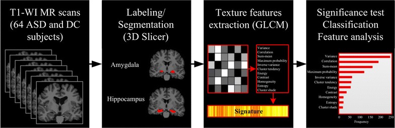
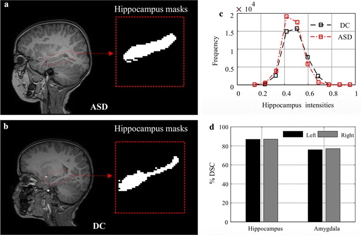
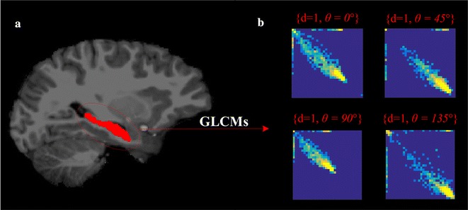
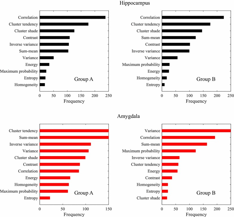
Background: Emerging evidence suggests the presence of neuroanatomical abnormalities in subjects with autism spectrum disorder (ASD). Identifying anatomical correlates could thus prove useful for the automated diagnosis of ASD. Radiomic analyses based on MRI texture features have shown a great potential for characterizing differences occurring from tissue heterogeneity, and for identifying abnormalities related to these differences. However, only a limited number of studies have investigated the link between image texture and ASD. This paper proposes the study of texture features based on grey level co-occurrence matrix (GLCM) as a means for characterizing differences between ASD and development control (DC) subjects. Our study uses 64 T1-weighted MRI scans acquired from two groups of subjects: 28 typical age range subjects 4-15 years old (14 ASD and 14 DC, age-matched), and 36 non-typical age range subjects 10-24 years old (20 ASD and 16 DC). GLCM matrices are computed from manually labeled hippocampus and amygdala regions, and then encoded as texture features by applying 11 standard Haralick quantifier functions. Significance tests are performed to identify texture differences between ASD and DC subjects. An analysis using SVM and random forest classifiers is then carried out to find the most discriminative features, and use these features for classifying ASD from DC subjects.

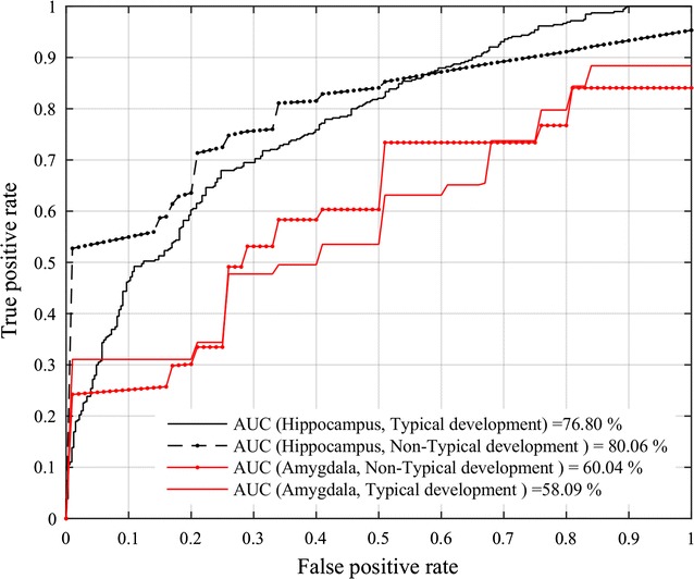
Results: Preliminary results show that all 11 features derived from the hippocampus (typical and non-typical age) and 4 features extracted from the amygdala (non-typical age) have significantly different distributions in ASD subjects compared to DC subjects, with a significance of p < 0.05 following Holm-Bonferroni correction. Features derived from hippocampal regions also demonstrate high discriminative power for differentiating between ASD and DC subjects, with classifier accuracy of 67.85%, sensitivity of 62.50%, specificity of 71.42%, and the area under the ROC curve (AUC) of 76.80% for age-matched subjects with typical age range.

Conclusions: Results demonstrate the potential of hippocampal texture features as a biomarker for the diagnosis and characterization of ASD.

Keywords: Autism spectrum disorder; Hippocampus; Radiomics.


Figures

Fig. 1
Fig. 1
Workflow of the proposed model. Data derived from T1-weighted MRI [scans reproduced with permission from the International Neuroimaging Data-Sharing Initiative (INDI), under the creative commons license (https://creativecommons.org/licenses/by-nc-sa/3.0/)]; manual labeling of hippocampus and amygdala regions; extraction of GLCM-based texture features from hippocampus and amygdala regions; identification of discriminative features for classifying ASD and DC subjects
Fig. 2
Fig. 2
Examples of hippocampus regions in ASD and DC subjects. a, b Examples of hippocampus segmentation masks for ASD and DC subjects (scans reproduced with permission from the International Neuroimaging Data-Sharing Initiative (INDI), under the creative commons license); c histogram of normalized intensities in MRI images of ASD and DC typical age range subjects; d dice similarity coefficient between the two expert labelings (left and right of hippocampus and amygdala regions, respectively)
Fig. 3
Fig. 3
GLCM computation. a Labeling of the hippocampus region in red (scans reproduced with permission from the International Neuroimaging Data-Sharing Initiative (INDI), under the creative commons license); b example of GLCMs corresponding to one offset and four different directions
Fig. 4
Fig. 4
ASD versus DC classification performance. Mean receiver operating characteristic (ROC) curve and AUC obtained by the SVM using the texture features derived from hippocampus (black curves) and amygdala (red curves) regions in typical (group A) and non-typical (group B) age range subjects
Fig. 5
Fig. 5
Dominant feature identification. (First row) Hippocampus-derived features; (second row) amygdala-derived features. Each bar represents the occurrence number of a feature in decision-tree root nodes (from 0 to 1000). Group A (left) and group B (right) contains typical and non-typical age range subjects, respectively

References

    1. Estes A, Zwaigenbaum L, Gu H, St John T, Paterson S, Elison JT, et al. Behavioral, cognitive, and adaptive development in infants with autism spectrum disorder in the first 2 years of life. J Neurodev Disord. 2015;7:24. doi: 10.1186/s11689-015-9117-6. - DOI - PMC - PubMed
    1. Minshew NJ, Payton JB. New perspectives in autism. Part I: the clinical spectrum of autism. Curr Probl Pediatr. 1988;18:561–610. - PubMed
    1. Tidmarsh L, Volkmar FR. Diagnosis and epidemiology of autism spectrum disorders. Can J Psychiatry Rev Can Psychiatr. 2003;48:517–525. doi: 10.1177/070674370304800803. - DOI - PubMed
    1. Stevens MC, Fein DA, Dunn M, Allen D, Waterhouse LH, Feinstein C, et al. Subgroups of children with autism by cluster analysis: a longitudinal examination. J Am Acad Child Adolesc Psychiatry. 2000;39:346–352. doi: 10.1097/00004583-200003000-00017. - DOI - PubMed
    1. Bedford R, Pickles A, Gliga T, Elsabbagh M, Charman T, Johnson MH, et al. Additive effects of social and non-social attention during infancy relate to later autism spectrum disorder. Dev Sci. 2014;17:612–620. doi: 10.1111/desc.12139. - DOI - PMC - PubMed

Publication types