. 2017 Aug 18;14(1):161.
doi: 10.1186/s12974-017-0933-3.

Age exacerbates microglial activation, oxidative stress, inflammatory and NOX2 gene expression, and delays functional recovery in a middle-aged rodent model of spinal cord injury

Affiliations

Age exacerbates microglial activation, oxidative stress, inflammatory and NOX2 gene expression, and delays functional recovery in a middle-aged rodent model of spinal cord injury

Ramona E von Leden et al. J Neuroinflammation. .

Abstract

Background: Spinal cord injury (SCI) among people over age 40 has been steadily increasing since the 1980s and is associated with worsened outcome than injuries in young people. Age-related increases in reactive oxygen species (ROS) are suggested to lead to chronic inflammation. The NADPH oxidase 2 (NOX2) enzyme is expressed by microglia and is a primary source of ROS. This study aimed to determine the effect of age on inflammation, oxidative damage, NOX2 gene expression, and functional performance with and without SCI in young adult (3 months) and middle-aged (12 months) male rats.

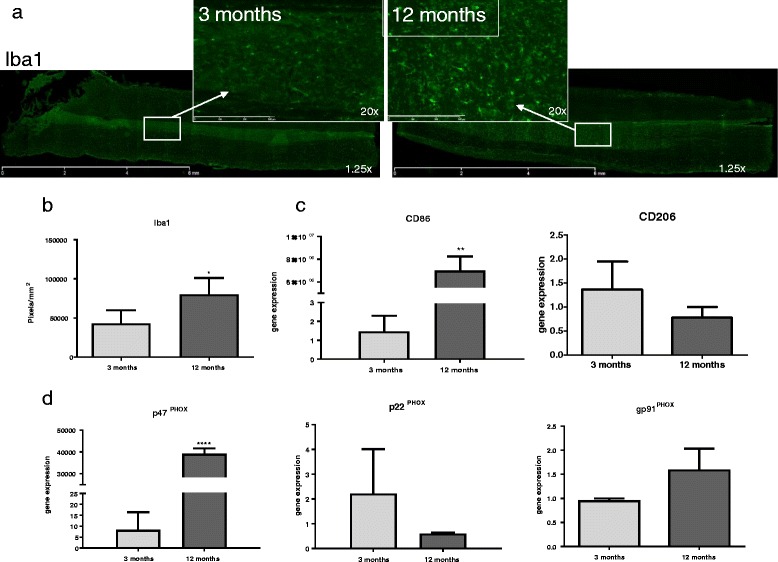
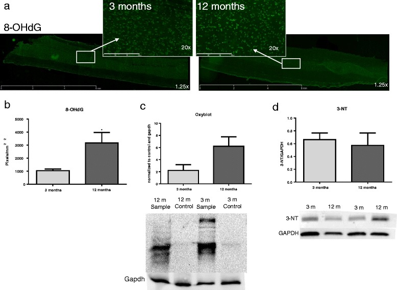
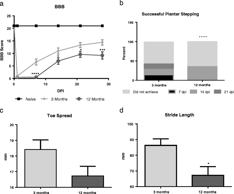
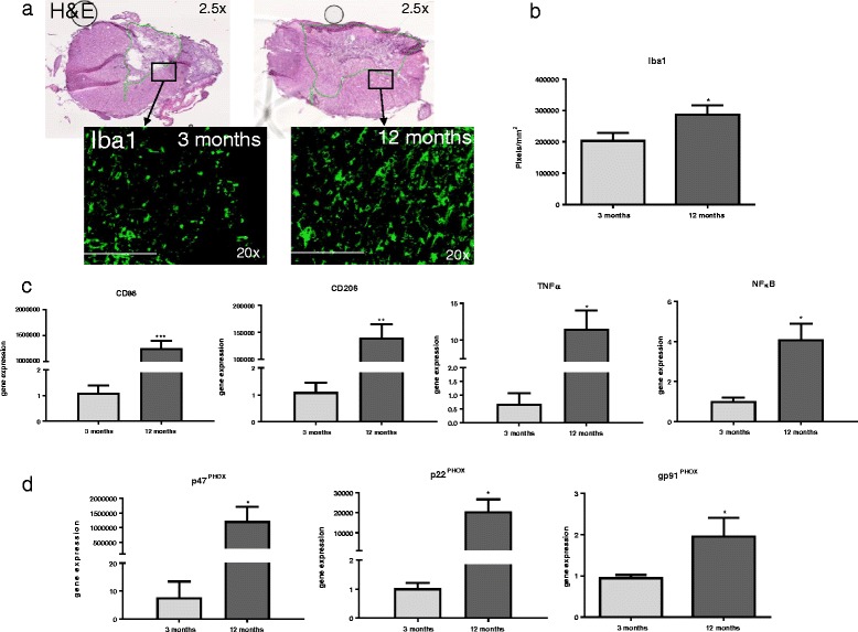
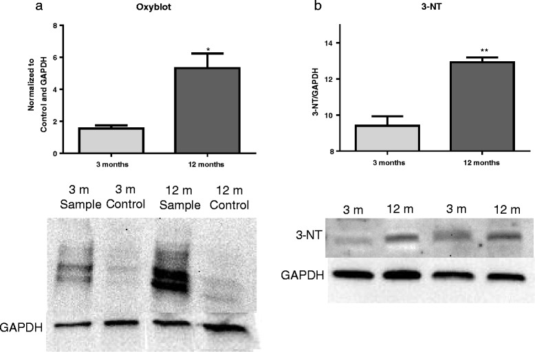
Methods: Young adult and middle-aged rats were assessed in two groups-naïve and moderate contusion SCI. Functional recovery was determined by weekly assessment with the Basso, Beattie, and Breshnahan general motor score (analyzed two-way ANOVA) and footprint analysis (analyzed by Chi-square analysis). Tissue was analyzed for markers of oxidative damage (8-OHdG, Oxyblot, and 3-NT), microglial-related inflammation (Iba1), NOX2 component (p47PHOX, p22PHOX, and gp91PHOX), and inflammatory (CD86, CD206, TNFα, and NFκB) gene expression (all analyzed by unpaired Student's t test).

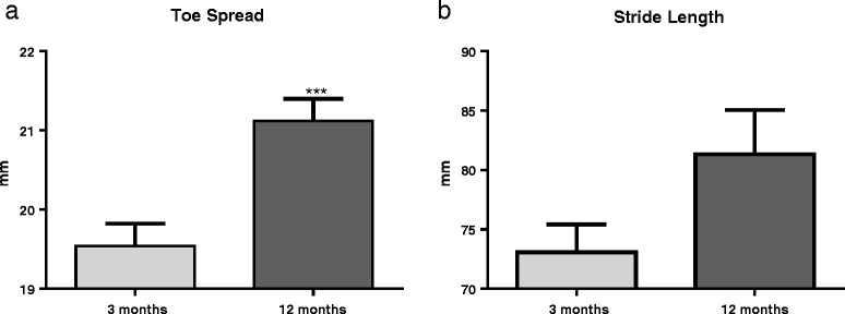
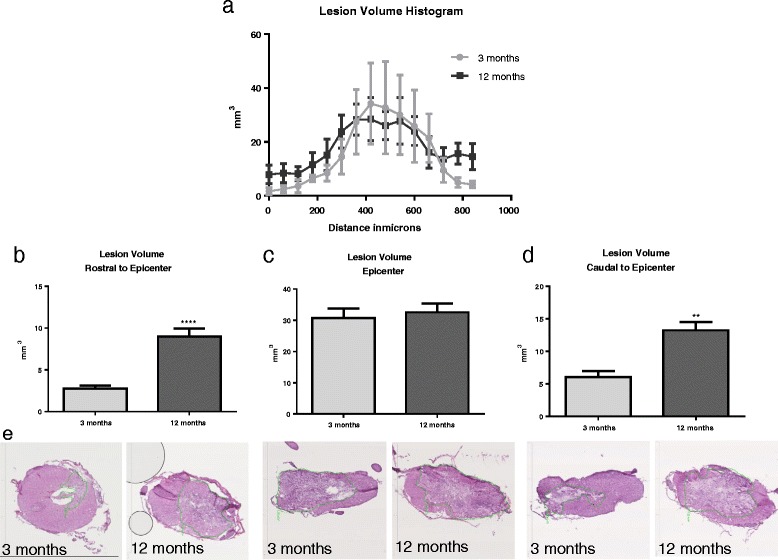
Results: In both naïve and injured aged rats, compared to young rats, tissue analysis revealed significant increases in 8-OHdG and Iba1, as well as inflammatory and NOX2 component gene expression. Further, injured aged rats showed greater lesion volume rostral and caudal to the injury epicenter. Finally, injured aged rats showed significantly reduced Basso-Beattie-Bresnahan (BBB) scores and stride length after SCI.

Conclusions: These results show that middle-aged rats demonstrate increased microglial activation, oxidative stress, and inflammatory gene expression, which may be related to elevated NOX2 expression, and contribute to worsened functional outcome following injury. These findings are essential to elucidating the mechanisms of age-related differences in response to SCI and developing age-appropriate therapeutics.

Keywords: Aging; Inflammation; Microglia; NOX2; Spinal cord injury.


Conflict of interest statement

Ethics approval

All animal procedures were approved by the Uniformed Services University IACUC and complied fully with the principles set forth in the “Guide for the Care and Use of Laboratory Animals” prepared by the Committee on Care and Use of Laboratory Animals of the Institute of Laboratory Resources, National Research Council (DHEW pub. No. (NIH) 85-23, 2985).

Consent for publication

Not Applicable.

Competing interests

The authors declare that they have no competing interests.

Publisher’s Note

Springer Nature remains neutral with regard to jurisdictional claims in published maps and institutional affiliations.

Figures

Fig. 1
Fig. 1
Uninjured aged animals show altered stepping pattern in footprint analysis. a Uninjured 12-month-old rats (n = 10) have significantly wider toe spread than 3-month-old rats (n = 12). b Uninjured 12-month old-rats show a trend toward increased stride length compared to 3-month-old rats, supporting toe spread findings. ***p < 0.001. Bars represent mean ± SEM
Fig. 2
Fig. 2
Microglia and NOX2 component activation and gene expression is altered with age. a Fluorescent microscope images of longitudinal sections of naïve 3- and 12-month-old rat spinal cords. Sections were stained with Iba-1 (green). Sections were imaged at 1.25× magnification and encompass an entire 10-mm section in the thoracic spinal cord. Inserts were taken at 20× magnification in the gray matter delineated by white boxes and arrows. b Immunostaining shows age-related differences in the microglial marker Iba1, showing significantly increased staining in 12- versus 3-month-old rats (size bar = 100 um). N = 4/group. c Comparative qPCR reveals significant increase in gene expression of the pro-inflammatory phenotype marker CD86. d Comparative RT-PCR reveals significant increase in p47PHOX gene expression in 12-month-aged rats compared to 3-month-aged rats. No significant difference was seen in gene expression of p22PHOX or gp91PHOX between the two groups. N = 4/group. *p < 0.05, **p < 0.005, ****p < 0.0005. Bars represent mean ± SEM
Fig. 3
Fig. 3
ROS production is increased with age. a Fluorescent microscope images of longitudinal sections of naïve 3- and 12-month-old rat spinal cords. Sections were stained with 8-OHdG (green). Sections were imaged at 1.25× magnification and encompass an entire 10-mm long section in the thoracic spinal cord. Inserts were taken at 20× magnification in the gray matter delineated by white boxes and arrows. b 8-OHdG stain shows significantly greater oxidative damage in 12 versus 3-month-old rats (size bar = 300 um). N = 4/group. c Oxyblot analysis shows a trend (p = 0.0946) toward increased protein oxidation in 12-month-aged rats compared to 3-months-aged rats. d Western blot analysis that shows protein expression of 3-NT, a marker of nitrosylated proteins which is a product of oxidative stress, is not significantly different between 12- and 3-month-aged rats. N = 4/group. *p < 0.05. Bars represent mean ± SEM
Fig. 4
Fig. 4
Aged animals show diminished motor function recovery after injury. a At 1 dpi, no difference in performance in BBB scores were observed in injured rats. At all other time points (7, 14, 21, and 28 dpi), 12-month-old injured rats (n = 17) showed significantly reduced BBB scores compared to 3-month-old rats (n = 18), indicating significantly impaired recovery to injury. b Aged animals show impaired ability to plantar step and complete footprint analysis task. Twelve-month-old rats (n = 17) show significantly diminished ability to plantar step in order to complete the footprint analysis task compared to 3-month-old rats (n = 14), indicating a diminished ability to take steps or strides. c At 28 dpi, toe spread analysis showed a trend in 12-month-old rats (n = 7) toward narrower toe spread compared to 3-month-old rats (n = 5). d 12-month-old rats (n = 7) showed significantly decreased stride length compared to 3-month-old rats (n = 5). *p < 0.05, **p < 0.005, ***p < 0.0005, ****p < 0.0001. Bars represent mean ± SEM
Fig. 5
Fig. 5
Aged rats have significantly great lesion volume rostral and caudal to the lesion. a Quantitative analysis of tissue stained with H&E reveals that at 30 dpi, 12-month-old rats show an increased lesion volume compared to 3-month-old rats both rostral and caudal to the injury epicenter, suggesting that aged rats have an increased volume and length of lesion compared to young. Lesion volume histogram shows the pattern of lesion spread from the epicenter. b Lesion volume rostral to the epicenter (0–180 μm) was significantly greater in 12-month-old rats compared to 3-month-old rats. c No significant difference was found in lesion volume at injury epicenter (360–540 μm). d Lesion volume caudal to the epicenter (720–900 μm) was significantly greater in 12-month-old aged rats compared to 3-month-old rats. e Photos depict sections of the spinal cord at each segment through the lesion from both 3- and 12-month-old rats. Rostral image is from a 120-μm section, epicenter image is from a 400-μm section, and caudal image is from a 820-μm section. Size bar = 3 mm. N = 6/group. **p < 0.005, ****p < 0.0001. Bars represent mean ± SEM
Fig. 6
Fig. 6
Microglial and NOX2 component activation and gene expression after spinal cord injury at 30 dpi. a 30 dpi H&E-stained images of 3 and 12-month-old injured spinal cord. Black boxes depict location of Iba1-stained quantification, in the closest intact tissue outside the central cavity of the injury. Black arrows point to corresponding images of immunofluorescent staining of Iba1. b Twelve-month-aged rats showed significantly greater staining in comparison to 3-month-aged rats at 30 dpi. N = 4/group. c Comparative RT-PCR reveals significant increase in gene expression of the M1 phenotype marker cd86, M2 phenotype marker cd206, and inflammatory cytokines TNFα and NFκB in 12-month-aged rats compared to that in 3-month-aged rats, suggesting an overall increase in microglial gene expression after injury with age. d Comparative RT-PCR of NOX2 components p47PHOX, p22PHOX, and gp91PHOX reveal a significant increase in gene expression of all three genes in 12-month-aged rats compared to 3-month-aged rats at 30 dpi. N = 4/group. *p < 0.05, **p < 0.005, ***p < 0.0005. Bars represent mean ± SEM
Fig. 7
Fig. 7
Aged rats show increased ROS production and oxidative stress at 30 dpi. a Oxyblot analysis reveals a significant increase in protein oxidation in 12-month-aged rats compared to 3-month-aged rats at 30 dpi. b Western blot analysis reveals a significant increase in 3-NT, a marker of nitrosylated proteins which is a product of oxidative stress, in 12-month-aged rats compared to that in 3-month-aged rats 30 dpi. N = 4/group. *p < 0.05, **p < 0.005. Bars represent mean ± SEM

References

    1. NSCISC . National Spinal Cord Injury Statistical Center, facts and figures at a glance. Birmingham: University of Alabama at Birmingham; 2016. p. 2016.
    1. Dumont RJ, Okonkwo DO, Verma S, Hurlbert RJ, Boulos PT, Ellegala DB, et al. Acute spinal cord injury, part I: pathophysiologic mechanisms. Clin Neuropharmacol. 2001;24:254–264. doi: 10.1097/00002826-200109000-00002. - DOI - PubMed
    1. Ronsyn MW, Berneman ZN, Van Tendeloo VFI, Jorens PG, Ponsaerts P. Can cell therapy heal a spinal cord injury? Spinal Cord. 2008;46:532–539. doi: 10.1038/sc.2008.13. - DOI - PubMed
    1. Jain NB, Ayers GD, Peterson EN, Harris MB, Morse L, O’Connor KC, et al. Traumatic spinal cord injury in the United States, 1993-2012. JAMA. 2015;313:2236–2243. doi: 10.1001/jama.2015.6250. - DOI - PMC - PubMed
    1. Lu T, Pan Y, Kao S-Y, Li C, Kohane I, Chan J, et al. Gene regulation and DNA damage in the ageing human brain. Nature. 2004;429:883–891. doi: 10.1038/nature02661. - DOI - PubMed

Publication types

MeSH terms