. 2017 Aug 18;10(1):39.
doi: 10.1186/s13041-017-0319-6.

Genetic deletion of mGlu2 metabotropic glutamate receptors improves the short-term outcome of cerebral transient focal ischemia

Affiliations

Genetic deletion of mGlu2 metabotropic glutamate receptors improves the short-term outcome of cerebral transient focal ischemia

Federica Mastroiacovo et al. Mol Brain. .

Abstract

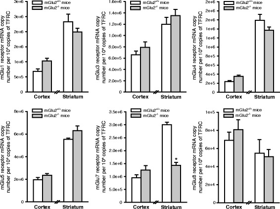
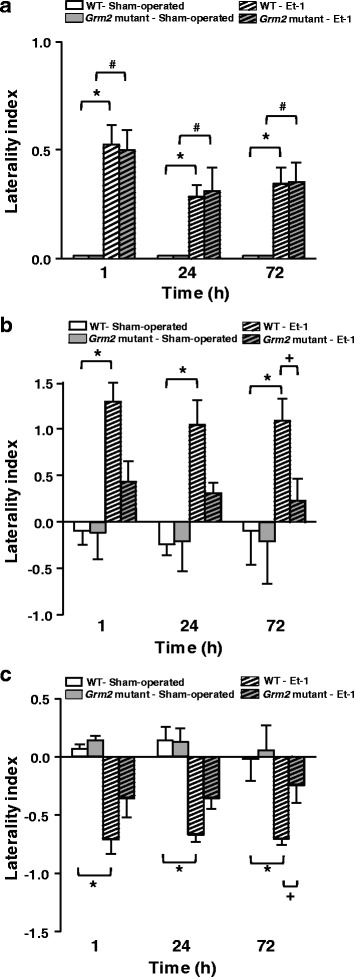
We have recently shown that pharmacological blockade of mGlu2 metabotropic glutamate receptors protects vulnerable neurons in the 4-vessel occlusion model of transient global ischemia, whereas receptor activation amplifies neuronal death. This raised the possibility that endogenous activation of mGlu2 receptors contributes to the pathophysiology of ischemic neuronal damage. Here, we examined this possibility using two models of transient focal ischemia: (i) the monofilament model of middle cerebral artery occlusion (MCAO) in mice, and (ii) the model based on intracerebral infusion of endothelin-1 (Et-1) in rats. Following transient MCAO, mGlu2 receptor knockout mice showed a significant reduction in infarct volume and an improved short-term behavioural outcome, as assessed by a neurological disability scale and the "grip test". Following Et-1 infusion, Grm2 gene mutated Hannover Wistar rats lacking mGlu2 receptors did not show changes in the overall infarct volume as compared to their wild-type counterparts, although they showed a reduced infarct area in the agranular insular cortex. Interestingly, however, mGlu2 receptor-deficient rats performed better than wild-type rats in the adhesive tape test, in which these rats did not show the laterality preference typically observed after focal ischemia. These findings support the hypothesis that activation of mGlu2 receptors is detrimental in the post-ischemic phase, and support the use of mGlu2 receptor antagonists in the experimental treatment of brain ischemia.

Keywords: Focal ischemia; Genetic deletion; Neurological score; Neuroprotection; mGlu2 receptor.


Conflict of interest statement

Ethics approval and consent to participate

Studies involving animals were performed in agreement with the National and International guidelines and regulations on animal care and use, and were approved by the Neuromed Institutional Animal Care and Use Committee. All efforts were made to minimize animal suffering and to reduce the number of animals used.

Consent for publication

Not applicable.

Competing interests

The author(s) declared no potential conflicts of interest with respect to the research, authorship, and/or publication of this article.

Publisher’s Note

Springer Nature remains neutral with regard to jurisdictional claims in published maps and institutional affiliations.

Figures

Fig. 1
Fig. 1
mGlu2 Receptor protein levels in the cerebral cortex of mGlu2+/+ mice subjected to transient MCAO. Representative images of Nissl staining in coronal sections of mice at 3, 6, 12 and 48 h after monofilament-induced transient MCAO are shown in (a). The boundaries of dissection of the core region and the neighboring cortical region used for Western blot analysis are shown in (b). Immunoblot analysis of mGlu2 receptors in the cortical region corresponding to the core and in the cortical regions neighboring the core (and the corresponding regions of the contralateral hemispheres and of non-ischemic mice) is shown in (c) and (d), respectively. Densitometric data were obtained as the sum of the two bands corresponding to monomers and dimers. Data are means + S.E.M. of 4–5 mice per group. *p < 0.05 as compared to all other groups (One-way ANOVA + Fisher’s PLSD; F2,10 = 9.26)
Fig. 2
Fig. 2
Reduced infarct volume in mGlu2−/− mice subjected to transient MCAO. Evans blue perfusion showing the absence of anatomical abnormalities of the MCA mGlu2−/− with respect to mGlu2+/+ mice (a, b). No difference in the reduction of cerebral blood flow in response to MCAO was found among mGlu2+/+ and mGlu2−/− mice. Nissl staining of mGlu2+/+ and mGlu2−/− mouse brains after transient MCAO. Mice were killed 48 h after reperfusion (c). Values of infarct volume (d) are means ± S.E.M. (n = 9 per group). + p < 0.05 (Student’s t-test; t16 = 1.75)
Fig. 3
Fig. 3
Improved short-term behavioral outcome in mGlu2−/− mice subjected to transient MCAO. Neurological deficit in mGlu2+/+ and mGlu2−/− mice subjected to transient MCAO (a). Bars are means + S.E.M. Friedman nonparametric ANOVA revealed significant effect of Time for mGlu2+/+ mice [Chi Sqr.(N=7, df=4) = 23.46, p = 0.0001] and for mGlu2−/− mice [Chi Sqr.(N=9, df=4) = 31.62, p = 0.000001]; () and (O) are Wilcoxon matched pairs comparisons between T0 (before ischemia) and 2, 24 and 48 h for mGlu2+/+ mice (n = 9) and mGlu2−/− mice (n = 11), respectively; + Kruskal-Wallis ANOVA and post-hoc Kruskal-Wallis test by ranks for difference between mGlu2+/+ and mGlu2−/− mice. Grip strength of ipsilateral and contralateral FL in mGlu2+/+ and mGlu2−/− mice subjected to transient MCAO (b). MCAO resulted into a significant reduction in muscular strength in both, ipsilateral and contralateral FLs, after ischemia compared to T0 in mGlu2+/+ (n = 7) and mGlu2−/− mice (n = 8) (p < 0.05, Dunnett’s t test). Three-way ANOVA for repeated measures (first factor GROUP with two levels: mGlu2+/+ and mGlu2−/− mice, second repeated measure factor SIDE with two levels: ipsilateral FL and contralateral FL, and third repeated measure factor TIME with four levels: T0, 2, 24, and 48 h) revealed significant effects of GROUP (F1,9 = 7.11, p = 0.026), SIDE (F1,9 = 6.69, p = 0.029), TIME (F3,27 = 21.16, p = 0.0000001), interactions SIDE x TIME (F3,27 = 3.83, p = 0.021) and GROUP x SIDE x TIME (F3,27 = 4.59, p = 0.01). & Comparisons between ipsilateral and contralateral FL in mGlu2+/+ mice (Fisher LSD test). + Comparisons between mGlu2+/+ and mGlu2−/− mice for the contralateral FL (Fisher LSD test)
Fig. 4
Fig. 4
Analysis of mGlu receptor subtypes in the cerebral cortex and striatum of mGlu2+/+ and mGlu2−/− mice. Quantitative PCR analysis of mGlu1, mGlu3, mGlu4, mGlu5, mGlu7 and mGlu8 receptors mRNA levels in mGlu2+/+ and mGlu2−/− mice. mRNA values, expressed as copy number, were normalized to TFRC (transferrin receptor protein-1) and are means + S.E.M. of 4–8 mice per group. * p < 0.05 (Student’s t test; t10 = 4.59)
Fig. 5
Fig. 5
No difference in the infarct volume between WT and Grm2 mutant rats subjected to MCAO. WT and Grm2 mutant chromatograms from Sanger sequencing (a); scheme design of a coronal brain section at bregma level + 0.2 mm [30] (b); representative images of brain Nissl staining in non-ischemic (sham-operated rat), WT and Grm2 mutant Han Wistar rats subjected to transient MCAO (c); total infarct volume and infarct volumes in the cerebral cortex and striatum are shown in (d), (e), and (f), respectively. Bars represent means + S.E.M. of 8 rats per group
Fig. 6
Fig. 6
Reduced infarct area in the agranular insular cortex of Grm2 mutant rats subjected to MCAO. Infarct areas of cortical sub-regions examined in one microscopic section (a) corresponding to the level of Et-1 infusion (AP = + 0.2 mm) are shown in (b-h). Bars are means + S.E.M. of 8 rats per group. * p < 0.05 (Student’s t test). S1FL, S1DZ, and S1ULP = forelimb region, dysgranular region, and upper lip region of the primary somatosensory cortex, respectively; S2 = secondary somatosensory cortex; GI/DI, AI = granular/dysgranular, and agranular insular cortex, respectively; Cl = claustrum
Fig. 7
Fig. 7
Improved motor asymmetry in Grm2 mutant Han Wistar rats subjected to transient MCAO. Asymmetry (expressed by laterality index, normalized to pre-ischemia values) in the PHR test is shown in (a). Kruskal-Wallis nonparametric ANOVA and Mann-Whitney comparisons between Et-1-infused and sham-operated rats: (*) for WT rats and (#) Grm2 mutant rats, respectively; Asymmetry in the preference and latency of removing the adhesive tapes in the AT test is shown in (b) and (c), respectively. Three-way GLM rANOVA (first factor ISCHEMIA with two levels: Et-1 and sham-operated, second factor GROUP with two levels: WT and Grm2 mutant rats, and third factor TIME with three levels: 1, 24 and 72 h) revealed a significant effect of factor ISCHEMIA for all rats, as follows: Preference (F(1,15) = 12.84, p = 0.003) and Latency (F(1,15) = 28.31, p = 0.00009). For ischemic rats (infused with Et-1), two-way rANOVA with factors GROUP (WT and Grm2 mutant rats) and TIME (1, 24 and 72 h) showed significant effect of the factor GROUP for both AT parameters, as follows: Preference (F(1,10) = 10.84, p = 0.008) and Latency (F(1,10) = 10.83, p = 0.008). * Fisher LSD comparisons between Et-1 and sham-operated WT rats and + between WT- Et-1 and Grm2 mutant-Et-1 rats. Number of rats: WT-sham-operated (n = 3); Grm2 mutant Han Wistar rats-sham-operated (n = 4); WT-Et-1 (n = 7); Grm2 mutant Han Wistar rats-Et-1 (n = 8). Significance was set at p < 0.05

Similar articles

Cited by

References

    1. Bruno V, Battaglia G, Copani A, D'Onofrio M, Di Iorio P, De Blasi A, et al. Metabotropic glutamate receptor subtypes as targets for neuroprotective drugs. J Cereb Blood Flow Metab. 2001;21(9):1013–1033. doi: 10.1097/00004647-200109000-00001. - DOI - PubMed
    1. Corti C, Battaglia G, Molinaro G, Riozzi B, Pittaluga A, Corsi M, et al. The use of knock-out mice unravels distinct roles for mGlu2 and mGlu3 metabotropic glutamate recptors in mechanisms of neurodegeneration/neuroprotection. J Neurosci. 2007;27(31):8297–8308. doi: 10.1523/JNEUROSCI.1889-07.2007. - DOI - PMC - PubMed
    1. Caraci F, Molinaro G, Battaglia G, Giuffrida ML, Riozzi B, Traficante A, et al. Targeting group II metabotropic glutamate (mGlu) receptors for the treatment of psychosis associated with Alzheimer’s disease: selective activation of mGlu2 receptors amplifies beta-amyloid toxicity in cultured neurons, whereas dual activation of mGlu2 and mGlu3 receptors is neuroprotective. Mol Pharmacol. 2011;79(3):618–626. doi: 10.1124/mol.110.067488. - DOI - PubMed
    1. Bruno V, Sureda FX, Storto M, Casabona G, Caruso A, Knopfel T, et al. The neuroprotective activity of group-II metabotropic glutamate receptors requires new protein synthesis and involves a glial-neuronal signaling. J Neurosci. 1997;17(6):1891–1897. - PMC - PubMed
    1. Bruno V, Battaglia G, Casabona G, Copani A, Caciagli F, Nicoletti F. Neuroprotection by glial metabotropic glutamate receptors is mediated by transforming growth factor-beta. J Neurosci. 1998;18(23):9594–9600. - PMC - PubMed

MeSH terms

Substances

LinkOut - more resources