. 2017 Oct 13;121(9):1058-1068.
doi: 10.1161/CIRCRESAHA.117.311607. Epub 2017 Aug 17.

Targeting miR-423-5p Reverses Exercise Training-Induced HCN4 Channel Remodeling and Sinus Bradycardia

Affiliations

Targeting miR-423-5p Reverses Exercise Training-Induced HCN4 Channel Remodeling and Sinus Bradycardia

Alicia D'Souza et al. Circ Res. .

Abstract

Rationale: Downregulation of the pacemaking ion channel, HCN4 (hyperpolarization-activated cyclic nucleotide gated channel 4), and the corresponding ionic current, If, underlies exercise training-induced sinus bradycardia in rodents. If this occurs in humans, it could explain the increased incidence of bradyarrhythmias in veteran athletes, and it will be important to understand the underlying processes.

Objective: To test the role of HCN4 in the training-induced bradycardia in human athletes and investigate the role of microRNAs (miRs) in the repression of HCN4.

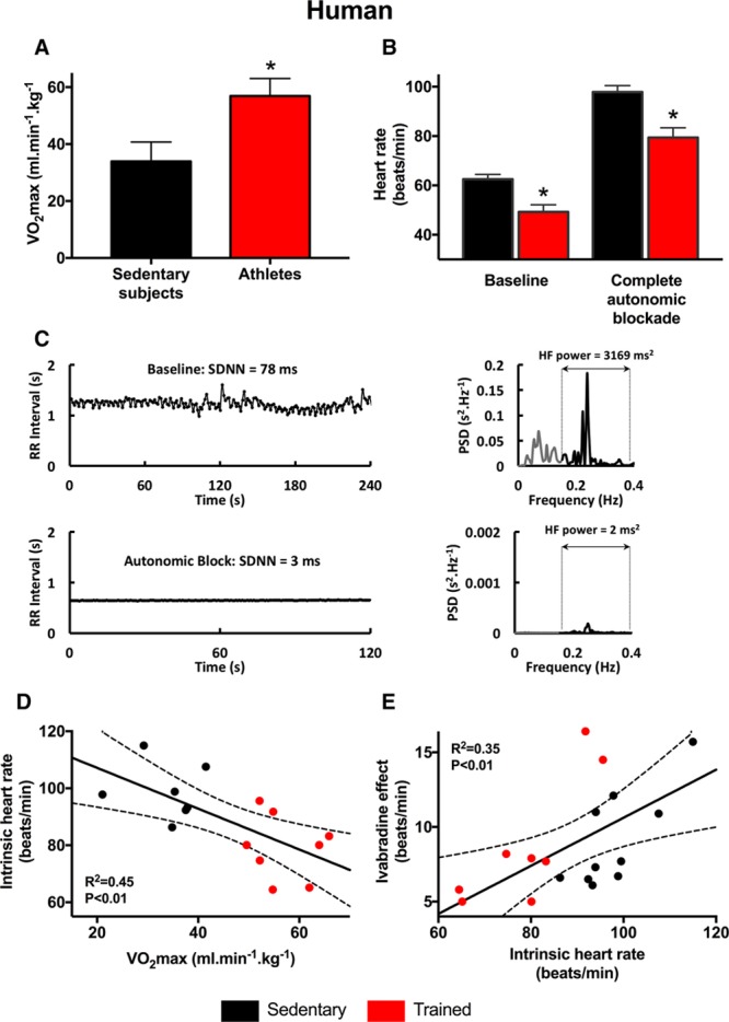
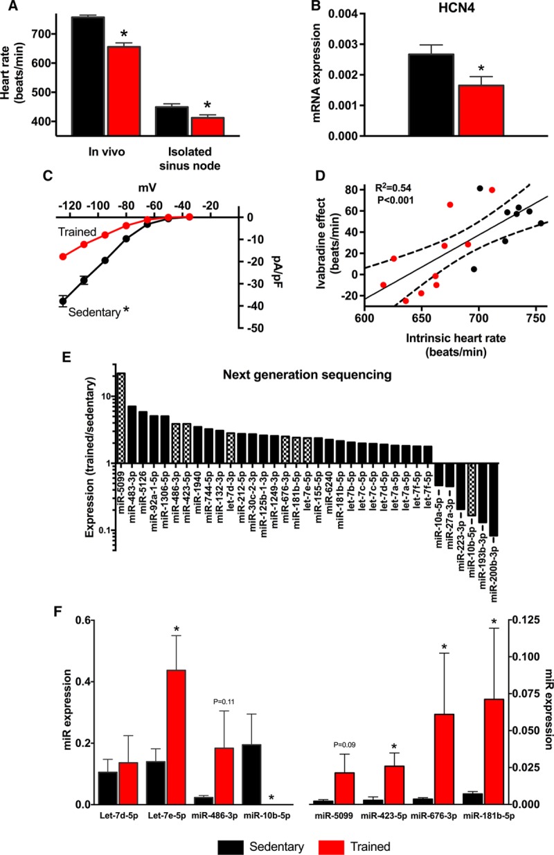
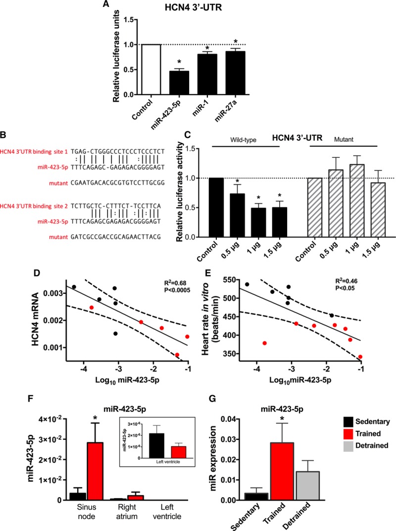
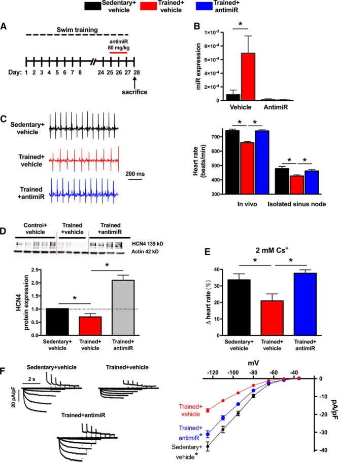
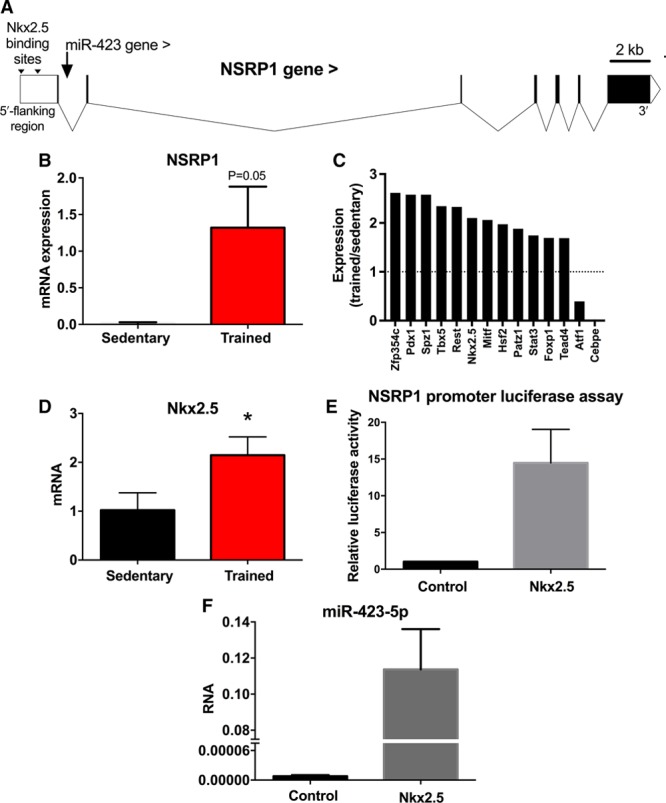
Methods and results: As in rodents, the intrinsic heart rate was significantly lower in human athletes than in nonathletes, and in all subjects, the rate-lowering effect of the HCN selective blocker, ivabradine, was significantly correlated with the intrinsic heart rate, consistent with HCN repression in athletes. Next-generation sequencing and quantitative real-time reverse transcription polymerase chain reaction showed remodeling of miRs in the sinus node of swim-trained mice. Computational predictions highlighted a prominent role for miR-423-5p. Interaction between miR-423-5p and HCN4 was confirmed by a dose-dependent reduction in HCN4 3'-untranslated region luciferase reporter activity on cotransfection with precursor miR-423-5p (abolished by mutation of predicted recognition elements). Knockdown of miR-423-5p with anti-miR-423-5p reversed training-induced bradycardia via rescue of HCN4 and If. Further experiments showed that in the sinus node of swim-trained mice, upregulation of miR-423-5p (intronic miR) and its host gene, NSRP1, is driven by an upregulation of the transcription factor Nkx2.5.

Conclusions: HCN remodeling likely occurs in human athletes, as well as in rodent models. miR-423-5p contributes to training-induced bradycardia by targeting HCN4. This work presents the first evidence of miR control of HCN4 and heart rate. miR-423-5p could be a therapeutic target for pathological sinus node dysfunction in veteran athletes.

Keywords: athletes; exercise training; ion channel remodeling; micro-RNAs; sinoatrial node; sinus bradycardia.


Figures

Figure 1.
Figure 1.
Evidence of a role for HCN4 (hyperpolarization-activated cyclic nucleotide gated channel 4) in the resting bradycardia in human athletes. A, VO2max of sedentary human subjects and human athletes (n=7/8). B, Heart rates measured under baseline conditions and after complete autonomic blockade of sedentary human subjects and human athletes (n=10/8). C, Complete autonomic blockade abolishes heart rate variability. Left, Examples traces of RR interval under baseline conditions and after complete autonomic blockade. Right, Spectral analyses of corresponding RR interval plots demonstrating near-abolition of high-frequency (HF) heart rate variability after autonomic blockade (note difference in y axis scale). D, Relationship between the intrinsic heart rate measured after complete autonomic blockade and VO2max in sedentary human subjects and human athletes (n=7/8). E, Relationship between ivabradine-induced decrease in heart rate and the intrinsic heart rate measured after complete autonomic blockade in sedentary human subjects and human athletes (n=10/8). In D and E, data fit by linear regression; best fit line, 95% confidence limits, and R2 and P values shown. PSD indicates power spectral density; and SDNN, standard deviation of normal to normal beats.
Figure 2.
Figure 2.
If remodeling in training-induced bradycardia is accompanied by dysregulation of miRs. A, Heart rates of sedentary and trained mice measured in vivo in conscious animals (n=5/9) and in vitro in isolated sinus node preparations (n=6/6). B, Expression of HCN4 (hyperpolarization-activated cyclic nucleotide gated channel 4) mRNA in sinus node of sedentary and trained mice (n=5/5). C, Current–voltage relationships for If recorded from single sinus node cells from sedentary (n=47 cells/5 mice) and trained (n=58 cells/4 mice) mice. D, Relationship between ivabradine-induced decrease in heart rate and the intrinsic heart rate measured in vivo after complete autonomic blockade in sedentary and trained mice (n=8/10). Data fit by linear regression; best fit line, 95% confidence limits, and R2 and P values are shown. E, Significant (as determined by DeSeq) training-induced changes in miR expression (measured by next generation sequencing) in sinus node of mice. Ratio of miR expression in trained mice to expression in sedentary mice shown on logarithmic scale. Hatched bars indicate significant differences after Benjamini–Hochberg false discovery rate (FDR) correction (P<0.05). Data obtained from 3 pooled RNA samples per group (n=2/2). F, Verification of training-induced changes in miRs by quantitative real-time reverse transcription polymerase chain reaction (qPCR). Expression shown in sedentary and trained mice (n=6/7–9). *Significant difference between sedentary and trained data (P<0.05).
Figure 3.
Figure 3.
HCN4 (hyperpolarization-activated cyclic nucleotide gated channel 4) is a target gene for miR-423-5p. A, Luciferase reporter assay showing post-transcriptional repression of HCN4 by miRs. H9C2 cells were cotransfected with precursor miR and 3′-UTR of HCN4 cloned into expression vector downstream of luciferase gene. Luciferase activity is shown 24 h after cotransfection with different miRs, including a control (scrambled) miR. n=3 batches of cells with 4 to 5 replicates/batch. B, Predicted miR-423-5p binding sites in HCN4 3′-UTR and corresponding sequence of mutant HCN4 3′-UTR tested. C, Luciferase reporter assay showing dose-dependent repression of HCN4 by miR-423-5p and loss of repression by mutation of HCN4 3′-UTR. Luciferase activity is shown 24 h after cotransfection with different amounts of wild-type or mutant miR-423-5p. n=3 batches of cells with 4 replicates/batch. D, Relationship between HCN4 mRNA and log miR-423-5p in sedentary and trained mice (n=5/5). E, Relationship between the heart rate measured in vitro in the isolated sinus node and log miR-423-5p in sedentary and trained mice (n=6/7). In D and E, data fit by linear regression; best fit line, 95% confidence limits, and R2 and P values are shown. F, Expression of miR-423-5p (measured by quantitative real-time reverse transcription polymerase chain reaction [qPCR]) in atrium and ventricle is low and unaltered by training. Expression shown in sinus node, right atrial muscle, and left ventricular muscle from sedentary and trained mice (n=5/5/5). Inset, data from left ventricular muscle at magnified scale. G, Expression of miR-423-5p (measured by qPCR) in sinus node is partially restored on detraining (right). Expression shown in sinus node of sedentary, trained, and detrained mice (n=5/9/5). *Significantly different from control/sedentary data (P<0.05).
Figure 4.
Figure 4.
Anti-miR to miR-423-5p reverses training-induced bradycardia and blunts HCN4 (hyperpolarization-activated cyclic nucleotide gated channel 4) channel remodeling. A, Time course of exercise training and anti-miR administration. B, Anti-miR abolishes training-induced upregulation of miR-423-5p in sinus node. miR-423-5p (determined by quantitative real-time reverse transcription polymerase chain reaction [qPCR]) in sinus node of vehicle- or anti–miR-treated sedentary and trained mice shown (n=6/5/5/5). C, Anti-miR reverses training-induced bradycardia. Representative ECG traces (recorded from conscious animals) from a vehicle-treated sedentary mouse, vehicle-treated trained mouse, and anti–miR-treated trained mouse shown on left and mean heart rates (measured in vivo and in isolated sinus node) on right (n=10/12/12 and 9/12/12). D, Anti-miR reverses training-induced downregulation of HCN4. Western blots using antibodies recognizing HCN4 and actin (housekeeper) proteins for sinus node from vehicle-treated sedentary, vehicle-treated trained, and anti-miR-treated trained mice shown as well as mean expression level of HCN4 protein (normalized to actin) in the 3 groups (n=5/4/5 with 3 independent replicates per mouse). E, Anti-miR reverses training-induced downregulation in contribution of If to pacemaking. Percentage decrease in heart rate (recorded from isolated sinus node preparations) on blocking If using 2 mmol/L Cs+ invehicle-treated sedentary, vehicle-treated trained, and anti-miR-treated trained mice shown (n=6/7/4). F, Anti-miR reverses training-induced downregulation in If. Representative If traces (normalized to cell capacitance) from vehicle-treated sedentary, vehicle-treated trained, and anti-miR-treated trained mice shown on left and mean current–voltage relationships for If from vehicle-treated sedentary (n=47 cells/5 animals), vehicle-treated trained (n=58 cells/4 animals), and anti-miR-treated trained (n=89 cells/5 animals) mice shown on right. *Significantly different (BE) or significantly different from trained+vehicle data (E; P<0.05).
Figure 5.
Figure 5.
Nkx2.5 regulation of miR-423-5p. A, map of NSRP1 gene (solid blocks show exons with introns in between) showing location of Nkx2.5 binding sites and intronic location of the miR-423 gene. B, Expression of NSRP1 mRNA in sinus node of sedentary and trained mice (n=8/8). C, Significant (P<0.05) training-induced changes in expression of transcription factor transcripts (measured by quantitative real-time reverse transcription polymerase chain reaction [qPCR]) in sinus node of mice. Ratio of mRNA expression in trained mice to expression in sedentary mice shown (n=6/8). D, Expression of Nkx2.5 mRNA in sinus node of sedentary and trained mice (n=8/8). E, Luciferase reporter assay showing activation of NSRP1 transcription by Nkx2.5. H9c2 cells were transfected with 2.1 kb of the 5′ flanking region of NSRP1 cloned into expression vector downstream of gene for luciferase. The cells were cotransfected with Nkx2.5; control cells were not cotransfected with Nkx2.5. Luciferase activity is shown 48 h after transfection. n=3 independent batches of cells with 4 replicates/batch. F, Upregulation of miR-423-5p by Nkx2.5. miR-423-5p expression is shown in H9c2 cells not transfected (control) or transfected with Nkx2.5.

Comment in

References

    1. Link MS, Homoud MK, Wang PJ, Estes NA., III Cardiac arrhythmias in the athlete: the evolving role of electrophysiology. Curr Sports Med Rep. 2002;1:75–85. - PubMed
    1. Northcote RJ, Canning GP, Ballantyne D. Electrocardiographic findings in male veteran endurance athletes. Br Heart J. 1989;61:155–160. - PMC - PubMed
    1. Northcote RJ, Rankin AC, Scullion R, Logan W. Is severe bradycardia in veteran athletes an indication for a permanent pacemaker? BMJ. 1989;298:231–232. - PMC - PubMed
    1. Baldesberger S, Bauersfeld U, Candinas R, Seifert B, Zuber M, Ritter M, Jenni R, Oechslin E, Luthi P, Scharf C, Marti B, Attenhofer Jost CH. Sinus node disease and arrhythmias in the long-term follow-up of former professional cyclists. Eur Heart J. 2008;29:71–78. doi: 10.1093/eurheartj/ehm555. - PubMed
    1. D’Souza A, Bucchi A, Johnsen AB, Logantha SJ, Monfredi O, Yanni J, Prehar S, Hart G, Cartwright E, Wisloff U, Dobryznski H, DiFrancesco D, Morris GM, Boyett MR. Exercise training reduces resting heart rate via downregulation of the funny channel HCN4. Nat Commun. 2014;5:3775. doi: 10.1038/ncomms4775. - PMC - PubMed

MeSH terms