. 2017 Sep 29;292(39):16300-16309.
doi: 10.1074/jbc.M117.804781. Epub 2017 Aug 15.

Distinct structural mechanisms determine substrate affinity and kinase activity of protein kinase Cα

Affiliations

Distinct structural mechanisms determine substrate affinity and kinase activity of protein kinase Cα

Sangbae Lee et al. J Biol Chem. .

Abstract

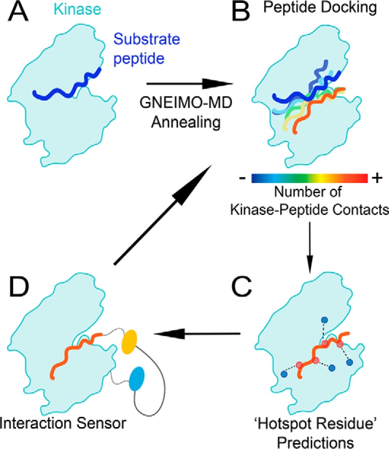
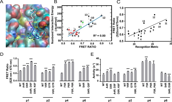
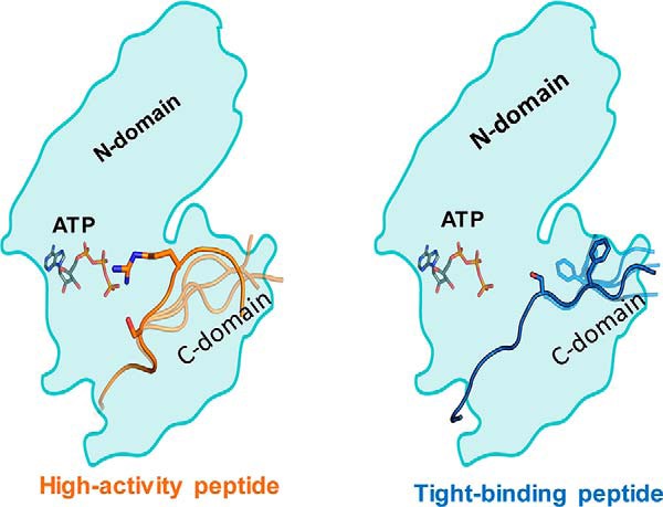
Protein kinase Cα (PKCα) belongs to the family of AGC kinases that phosphorylate multiple peptide substrates. Although the consensus sequence motif has been identified and used to explain substrate specificity for PKCα, it does not inform the structural basis of substrate-binding and kinase activity for diverse substrates phosphorylated by this kinase. The transient, dynamic, and unstructured nature of this protein-protein interaction has limited structural mapping of kinase-substrate interfaces. Here, using multiscale MD simulation-based predictions and FRET sensor-based experiments, we investigated the conformational dynamics of the kinase-substrate interface. We found that the binding strength of the kinase-substrate interaction is primarily determined by long-range columbic interactions between basic (Arg/Lys) residues located N-terminally to the phosphorylated Ser/Thr residues in the substrate and by an acidic patch in the kinase catalytic domain. Kinase activity stemmed from conformational flexibility in the region C-terminal to the phosphorylated Ser/Thr residues. Flexibility of the substrate-kinase interaction enabled an Arg/Lys two to three amino acids C-terminal to the phosphorylated Ser/Thr to prime a catalytically active conformation, facilitating phosphoryl transfer to the substrate. The structural mechanisms determining substrate binding and catalytic activity formed the basis of diverse binding affinities and kinase activities of PKCα for 14 substrates with varying degrees of sequence conservation. Our findings provide insight into the dynamic properties of the kinase-substrate interaction that govern substrate binding and turnover. Moreover, this study establishes a modeling and experimental method to elucidate the structural dynamics underlying substrate selectivity among eukaryotic kinases.

Keywords: GNEIMO; binding affinity; conformational change; fluorescence resonance energy transfer (FRET); molecular dynamics; multiscale molecular dynamics; peptide interaction; protein conformation; protein kinase C (PKC); protein phosphorylation.


Conflict of interest statement

The authors declare that they have no conflicts of interest with the contents of this article

Figures

Figure 1.
Figure 1.
Combining computational and experimental approaches to dissect the kinase–substrate interaction. A, PKC–substrate interaction is homology modeled from the PKCι–Par3 interaction (PDB ID 5HI1). B, GNEIMO torsional MD is used to obtain a kinase–substrate conformational ensemble. C, structural analysis is used to identify hot spot residues and motifs within the kinase and substrate that contribute to binding and catalytic activity. D, SPASM FRET sensors are used to test GNEIMO-MD predictions using site-directed mutagenesis. Experimental results are used to validate GNEIMO-MD predictions and refine computational methods.
Figure 2.
Figure 2.
Electrostatic interactions between residues in the N terminus of the peptide substrate and the kinase catalytic domain are major determinants of binding strength. A, predicted binding site of the tight binding peptide substrate p12 in PKCα. The backbone atoms of the residues before the Ser that gets phosphorylated in the substrate p12 are shown in blue and the residues after Ser are shown in white. The residues in the peptide substrate are shown in sticks. The residues in PKCα that make strong contact with the peptide are shown in ball and sticks. B, calculated average interaction energy of the N terminus of the peptide substrates with PKCα versus FRET intensity ratio. For clarity, the weak, medium, and strong binding peptide substrates are shown in red, green, and black spheres, respectively. The error bars for FRET measurement and the non-bond interaction energy represent the mean ± S.E. and the standard deviation, respectively. C, FRET ratio of SPASM sensors show a linear correlation with a recognition metric derived by Nishikawa et al. (14). D, site-directed mutagenesis of residues in peptides p1, p2, p4, and p6 identified from the MD simulation analysis to improve the binding affinity. Mutagenesis of N-terminal residues to Arg enhances kinase-substrate interaction as measured by FRET ratio. E, kinase activity of mutant peptide substrates shown in D. Mutagenesis of N-terminal residues to Arg marginally increase kinase specific activity compared with wild-type counterparts. The FRET results are expressed as mean ± S.E. of three independent experiments performed in triplicate (n ≥ 9). *, p < 0.05; **, p < 0.005; ***, p < 0.0005; ****, p < 0.0001.
Figure 3.
Figure 3.
High activity peptides populate catalytically favorable conformations when bound to the kinase. A and B, snapshots from MD trajectories showing the plausible catalytic conformation of the high activity peptide p2 (A) and low activity peptide p12 (B), bound to PKCα. These snapshots were selected based on the shortest distance between the Ser in the substrate to Asp466 that abstracts the proton from Ser and also to the γ-PO4 group of the ATP. C, population distribution of various peptide conformations from three 100-ns all-atom MD trajectories for peptide substrates p2 and p12. The MD ensemble was clustered by two distances. The distance between ATP(γP) and S/T(OG) of the substrate peptide and between D466(OD) and S/T(OG). The cross marks in these two figures are the conformations for which these distances are shown in A and B. D, population density of the catalytic conformations as captured in the MD simulations. We used a cutoff of 4.2 Å in the ATP(γP) and S/T(OG) distance and 4.1 Å in the D466(OD) and S/T(OG) distance to calculate this population density. E, rotamer flip change in the χ1 angle of the pSer/Thr in peptide substrates as seen from crystal structures before (PDB code 4XW5, PKA) and after (PDB code 4IAK, PKA) the phosphoryl transfer. F, all-atom MD simulations after the PO4 transfer was done showed a rotamer flip in pSer in the good activity peptide substrate p1 after the PO4 transfer to the pSer in the peptide. The panel shows the most populated rotamer of Ser/Thr in the simulations of the peptide p1 before and after the PO4 transfer. The peptide backbone is colored in yellow and the Ser and pSer are shown in sticks.
Figure 4.
Figure 4.
Arginine positioned two or three residues C-terminal to the phosphorylated serine in the peptide substrate is necessary and sufficient for high kinase activity. A, plot of the time variation of the distance between the γ-PO4 group of ATP and the OG atom in Ser in peptide substrates p2 and p12 shown in black lines and the distance between residue at position 11 to the γ-PO4 group of ATP shown in red lines. B, representative snapshot from the MD simulations showing the close distance between the side chain Arg11 and Ser9 of the peptide to the γ-PO4 group of ATP. C, kinase activity measured for mutants of position 11 in peptide substrates p9, p10, p12, and p2 to show the role of Arg in position 11 of the peptides to improve the activity. Arg at positions 11 or 12 is necessary for high kinase activity. D, FRET ratio of peptide substrates. Results are expressed as mean ± S.E. of three independent experiments performed in triplicate (n ≥ 9). *, p < 0.05; **, p < 0.005; ***, p < 0.0005; ****, p < 0.0001.
Figure 5.
Figure 5.
Distinct mechanisms of kinase activity and substrate-binding strengths. Left, high activity peptides display greater conformational flexibility in the domain C-terminal to the phosphorylated Ser/Thr (C-domain). An Arg/Lys residue 2–3 amino acids C-terminal to the phosphorylated Ser/Thr threads the formation of a catalytically active conformation. Right, tight binding peptides have several Arg/Lys residues N-terminal to the phosphorylated Ser/Thr that undergo strong columbic interactions with an acidic patch in the kinase catalytic domain.

Similar articles

Cited by

References

    1. Rust H. L., and Thompson P. R. (2011) Kinase consensus sequences: a breeding ground for crosstalk. ACS Chem. Biol. 6, 881–892 - PMC - PubMed
    1. Zaidel-Bar R., Itzkovitz S., Ma'ayan A., Iyengar R., and Geiger B. (2007) Functional atlas of the integrin adhesome. Nat. Cell Biol. 9, 858–867 - PMC - PubMed
    1. Sommese R. F., and Sivaramakrishnan S. (2016) Substrate affinity differentially influences protein kinase C regulation and inhibitor potency. J. Biol. Chem. 291, 21963–21970 - PMC - PubMed
    1. Endicott J. A., Noble M. E., and Johnson L. N. (2012) The structural basis of control of eukaryotic protein kinases. Annu. Rev. Biochem. 81, 587–613 - PubMed
    1. de Oliveira P. S., Ferraz F. A., Pena D. A., Pramio D. T., Morais F. A., and Schechtman D. (2016) Revisting protein kinase-substrate interactions: towards therapeutic development. Sci. Signal. 9, re3. - PubMed

Publication types

MeSH terms

LinkOut - more resources