. 2017 Sep 29;292(39):16333-16350.
doi: 10.1074/jbc.M117.802025. Epub 2017 Aug 18.

Dsc E3 ligase localization to the Golgi requires the ATPase Cdc48 and cofactor Ufd1 for activation of sterol regulatory element-binding protein in fission yeast

Affiliations

Dsc E3 ligase localization to the Golgi requires the ATPase Cdc48 and cofactor Ufd1 for activation of sterol regulatory element-binding protein in fission yeast

Risa Burr et al. J Biol Chem. .

Abstract

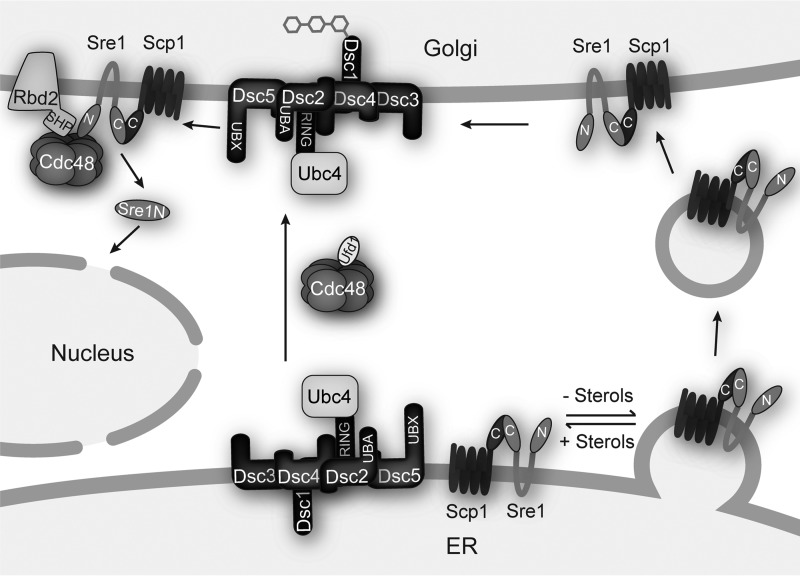
Sterol regulatory element-binding proteins (SREBPs) in the fission yeast Schizosaccharomyces pombe regulate lipid homeostasis and the hypoxic response under conditions of low sterol or oxygen availability. SREBPs are cleaved in the Golgi through the combined action of the Dsc E3 ligase complex, the rhomboid protease Rbd2, and the essential ATPases associated with diverse cellular activities (AAA+) ATPase Cdc48. The soluble SREBP N-terminal transcription factor domain is then released into the cytosol to enter the nucleus and regulate gene expression. Previously, we reported that Cdc48 binding to Rbd2 is required for Rbd2-mediated SREBP cleavage. Here, using affinity chromatography and mass spectrometry experiments, we identified Cdc48-binding proteins in S. pombe, generating a list of many previously unknown potential Cdc48-binding partners. We show that the established Cdc48 cofactor Ufd1 is required for SREBP cleavage but does not interact with the Cdc48-Rbd2 complex. Cdc48-Ufd1 is instead required at a step prior to Rbd2 function, during Golgi localization of the Dsc E3 ligase complex. Together, these findings demonstrate that two distinct Cdc48 complexes, Cdc48-Ufd1 and Cdc48-Rbd2, are required for SREBP activation and low-oxygen adaptation in S. pombe.

Keywords: ATPases associated with diverse cellular activities (AAA); Cdc48; E3 ubiquitin ligase; SREBP; Schizosaccharomyces pombe; VCP; hypoxia; membrane transport; p97; transcription regulation.


Conflict of interest statement

The authors declare that they have no conflicts of interest with the contents of this article

Figures

Figure 1.
Figure 1.
Identification of cdc48 alleles. A, flow chart of results from two MMS mutagenesis screens (0.024 and 0.012% (w/v)). Red numbers denote the number of isolates in each category. Numbers in parentheses are the number of isolates with mutations confirmed by sequencing (see supplemental Table S1 for specific mutations). Aside from the first box, all numbers are pooled from the two MMS dosages. B, comparison of gene-coding sequence size with number of isolated mutants in MMS mutagenesis screen. Best-fit line calculated against non-essential genes (filled markers), R2 = 0.87. C, table of remade cdc48 alleles showing amino acid changes in S. pombe, corresponding to homologous amino acids in humans and corresponding allele from our previous report (4). D, line diagram of S. pombe cdc48, where the orange box indicates the N domain to which the majority of cofactors bind. The blue box indicates the minor ATPase D1 domain required for hexamer formation. The green box indicates the major ATPase D2 domain required for force generation. Yellow boxes in both domains indicate sequences essential for ATP binding and hydrolysis. The gray box indicates the C domain to which a minority of cofactors bind. Red asterisks indicate the locations of our cdc48 point mutations.
Figure 2.
Figure 2.
SREBP cleavage requires cdc48. A, wild-type cells or the indicated mutants (5000 cells) were grown on rich medium plus or minus CoCl2 for 2 or 10 days, respectively. B, Western blots, probed with monoclonal anti-Sre1 IgG (5B4) and polyclonal anti-Dsc5 IgG (for loading), of lysates treated with alkaline phosphatase for 1 h from wild-type cells and the indicated cdc48 mutants grown for 0 or 4 h in the absence of oxygen. P and N denote precursor and cleaved N-terminal transcription factor forms, respectively. Asterisk denotes non-specific band. C, quantification of Sre1 N terminus from B of two biological replicates each denoted by different marker symbols. The quantity of Sre1N was normalized to Dsc5 for loading and then to the WT 4-h sample (lane 2) for comparison between blots. D, Western blots, probed with monoclonal anti-FLAG M2 and polyclonal anti-Dsc5 IgG (for loading), of lysates treated with alkaline phosphatase for 1 h from wild-type cells and the indicated cdc48 mutants containing a plasmid expressing sre2-MS (+) or the empty vector (−) grown in the presence of oxygen. P and N denote precursor and cleaved N-terminal transcription factor forms, respectively. E, quantification of Sre2-MS cleavage from D of three biological replicates each denoted by different marker symbols. The quantity of precursor and N terminus were normalized first to empty vector and then to Dsc5 for loading. Percent cleavage in each sample was calculated by dividing the normalized quantity of N terminus by the total signal (N+P). Error bars are 1 S.D. (**, p < 0.01 versus WT by two-tailed Student's t test).
Figure 3.
Figure 3.
cdc48 mutants have minimal effect on Cdc48 expression or growth rate. A, Western blottings, probed with polyclonal anti-Cdc48 IgG and polyclonal anti-Dsc5 IgG (for loading), of lysates from wild-type cells and the indicated cdc48 mutants. B, quantification of A from three biological replicates, each indicated by a different marker shape, normalized for loading to Dsc5 and then normalized to wild-type cells for comparison among blots. Error bars are 1 S.D. (*, p < 0.05; **, p < 0.01 versus WT by two-tailed Student's t test). C, indicated strains were grown in liquid culture for 12 h. Cell density was measured by absorbance at 600 nm every 3 h. Doubling times were calculated using 3- and 9-h optical density readings using the formula: doubling time = (culture time × log(2))/log(final OD) − log(initial OD). Doubling times are displayed as mean ± S.D. from four biological replicates as indicated by marker shape. (*, p < 0.05; **, p < 0.01 versus WT by two-tailed Student's t test).
Figure 4.
Figure 4.
Identification of Cdc48-binding proteins in S. pombe. A, Western blottings, probed with monoclonal anti-Sre1 IgG (5B4) and polyclonal anti-Dsc5 IgG (for loading), of lysates treated with alkaline phosphatase for 1 h from wild-type cells, cdc48-5xFLAG (cdc48-F) cells, or sre1Δ cells grown for 0 or 4 h in the absence of oxygen. P and N denote precursor and cleaved N-terminal transcription factor forms, respectively. Asterisks denote non-specific bands. B, Cdc48 was immunoprecipitated (IP) from wild-type or cdc48-5xFLAG cells using monoclonal anti-FLAG M2 IgG as described under “Experimental procedures.” Input, unbound, and 10-fold enriched bound fractions were analyzed by Western blotting using polyclonal anti-Cdc48 IgG. The blot is representative of four biological replicates. C, volcano plot of all proteins identified during TMT mass spectrometry of Cdc48–5xFLAG bound proteins. Points in red are proteins with enrichment >2 S.D. from the mean in the Cdc48–5xFLAG versus wild-type samples. p values were calculated using two-way ANOVA with quantile normalization for three biological replicates.
Figure 5.
Figure 5.
Ufd1 is a Cdc48 cofactor required for SREBP cleavage. A, table of ufd1 alleles used in this study. B, wild-type cells or the indicated mutants (5000 cells) were grown on rich medium plus or minus CoCl2 for 10 days at 30 °C. C, indicated strains were grown in liquid culture for 12 h at 30 °C. Cell density was measured by absorbance at 600 nm every 3 h. Doubling times were calculated using 3- and 9-h optical density readings using the formula: doubling time = (culture time × log(2))/log(final OD) −log(initial OD). Doubling times are displayed as mean ± S.D. for three biological replicates as indicated by marker shape. (**, p < 0.01 versus WT by two-tailed Student's t test). D, Western blottings, probed with monoclonal anti-Sre1 IgG (5B4) and polyclonal anti-Dsc5 IgG (for loading), of lysates treated with alkaline phosphatase for 1 h from wild-type cells, sre1Δ, or the indicated ufd1 mutants grown for 0 or 4 h in the absence of oxygen. P and N denote precursor and cleaved N-terminal transcription factor forms, respectively. Asterisk denotes non-specific band. E, quantification of Sre1 N terminus from D of two biological replicates each denoted by different marker symbols. The quantity of Sre1N was normalized to Dsc5 for loading and then to the WT 4 h sample (lane 2) for comparison between blots. F, Western blottings, probed with monoclonal anti-FLAG M2 and polyclonal anti-Dsc5 IgG (for loading), of lysates treated with alkaline phosphatase for 1 h from wild-type cells and the indicated ufd1 mutants containing a plasmid expressing sre2-MS (+) or the empty vector (−) grown in the presence of oxygen. P and N denote precursor and cleaved N-terminal transcription factor forms, respectively. The blot is representative of three biological replicates. G, quantification of Sre2-MS cleavage from F of three biological replicates each denoted by different marker symbols. The quantity of precursor and N terminus were normalized first to empty vector and then to Dsc5 for loading. Percent cleavage in each sample was calculated by dividing the normalized quantity of N terminus by the total signal (N+P). Error bars are 1 S.D. (*, p < 0.05; **, p < 0.01 versus WT by two-tailed Student's t test).
Figure 6.
Figure 6.
Cdc48-Ufd1 complex formation is altered in ufd1 and cdc48 mutants. A and C, Nonidet P-40-solubilized membrane protein was prepared from WT (−), ufd1–13xmyc (WT), or the indicated ufd1 mutant cells, and Ufd1–13xMyc was immunoprecipitated (IP) with anti-Myc monoclonal IgG as described under “Experimental procedures.” Equal quantities of input, unbound, and bound fractions were analyzed by immunoblotting using polyclonal antibodies against Cdc48 and Myc. B and D, quantification of Cdc48 pulldown from A or C of four (A) or five (C) biological replicates each denoted by different marker symbols. The quantity of Cdc48 in the bound fractions was normalized to the quantity of Myc in the bound fractions and then to the WT sample for comparison between blots. Error bars are 1 S.D. (*, p < 0.05; **, p < 0.01 versus WT by two-tailed Student's t test).
Figure 7.
Figure 7.
SREBP cleavage requires Cdc48-Ufd1 during Dsc E3 ligase Golgi localization. A, recombinant GST-HA-V5 control (GST), GST-Dsc5 UBX (Dsc5(323–425), UBX), and GST-Rbd2 C terminus (Rbd2(200–251), SHP) were bound to GST magnetic beads and incubated with S. pombe cytosol from wild-type cells (−) or ufd1–13xmyc cells (+). Input, unbound, and 10-fold enriched bound fractions were probed with monoclonal anti-Myc IgG, polyclonal anti-Cdc48 IgG, and monoclonal anti-GST IgG. The blot is representative of five replicates. B and D, Western blottings, probed with polyclonal anti-Sre2 IgG and polyclonal anti-Dsc5 IgG (for loading), of lysates treated with alkaline phosphatase for 1 h from wild-type, dsc1Δ (1Δ), or the indicated cdc48 (B) or ufd1 (D) mutant cells. rbd2 (+) or rbd2Δ (−) genotype is indicated. P and N denote precursor and cleaved N-terminal transcription factor forms, respectively. C and E, quantification of Sre2 precursor from B and D of three (B) or four (D) biological replicates each denoted by different marker symbols. The quantity of precursor was normalized to Dsc5 for loading and then to WT rbd2+ (lane 1) for comparison between blots. Error bars are 1 S.D. (*, p < 0.05; **, p < 0.01 versus WT rbd2+ by two-tailed Student's t test). Note, C, lane 3, has only two replicates, and therefore p values could not be calculated. F, Western blotting, probed with polyclonal anti-Dsc1 IgG, of Nonidet P-40-solubilized membrane protein from wild-type, dsc2Δ, dsc5ΔUBXUBX), rbd2Δ, or the indicated cdc48 or ufd1 mutant cells. M and I indicate mature and intermediate glycosylated forms, respectively. The blot is representative of three biological replicates. G, quantification of Dsc1 from F of three biological replicates each denoted by different marker symbols. The quantity of the mature form was divided by total Dsc1 signal for percent mature, allowing comparison between lanes and blots. Error bars are 1 S.D. (*, p < 0.05; **, p < 0.01 versus WT by two-tailed Student's t test).
Figure 8.
Figure 8.
SREBP activation mechanism in S. pombe. Model outlining the steps of SREBP transport and cleavage in the Golgi, incorporating the two roles of Cdc48 at Dsc E3 ligase Golgi localization and Rbd2.

Similar articles

Cited by

References

    1. Hughes A. L., Todd B. L., and Espenshade P. J. (2005) SREBP pathway responds to sterols and functions as an oxygen sensor in fission yeast. Cell 120, 831–842 - PubMed
    1. Kwon E. J., Laderoute A., Chatfield-Reed K., Vachon L., Karagiannis J., and Chua G. (2012) Deciphering the transcriptional-regulatory network of flocculation in Schizosaccharomyces pombe. PLoS Genet. 8, e1003104. - PMC - PubMed
    1. Stewart E. V., Nwosu C. C., Tong Z., Roguev A., Cummins T. D., Kim D. U., Hayles J., Park H. O., Hoe K. L., Powell D. W., Krogan N. J., and Espenshade P. J. (2011) Yeast SREBP cleavage activation requires the Golgi Dsc E3 ligase complex. Mol. Cell 42, 160–171 - PMC - PubMed
    1. Stewart E. V., Lloyd S. J., Burg J. S., Nwosu C. C., Lintner R. E., Daza R., Russ C., Ponchner K., Nusbaum C., and Espenshade P. J. (2012) Yeast sterol regulatory element-binding protein (SREBP) cleavage requires Cdc48 and Dsc5, a ubiquitin regulatory X domain-containing subunit of the Golgi Dsc E3 ligase. J. Biol. Chem. 287, 672–681 - PMC - PubMed
    1. Burr R., Stewart E. V., Shao W., Zhao S., Hannibal-Bach H. K., Ejsing C. S., and Espenshade P. J. (2016) Mga2 transcription factor regulates an oxygen-responsive lipid homeostasis pathway in fission yeast. J. Biol. Chem. 291, 12171–12183 - PMC - PubMed

MeSH terms

Substances

LinkOut - more resources